. 2020 Apr 16;524(4):923-928.
doi: 10.1016/j.bbrc.2020.02.016. Epub 2020 Feb 11.

Targeting Hsc70-based autophagy to eliminate amyloid β oligomers

Affiliations

Targeting Hsc70-based autophagy to eliminate amyloid β oligomers

Juan Dou et al. Biochem Biophys Res Commun. .

Abstract

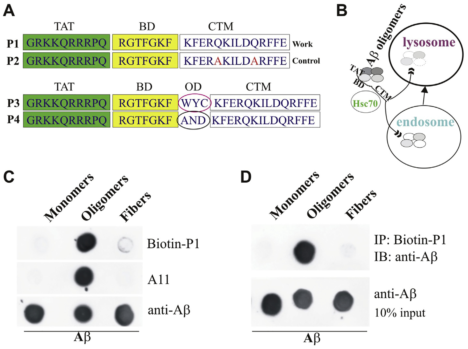
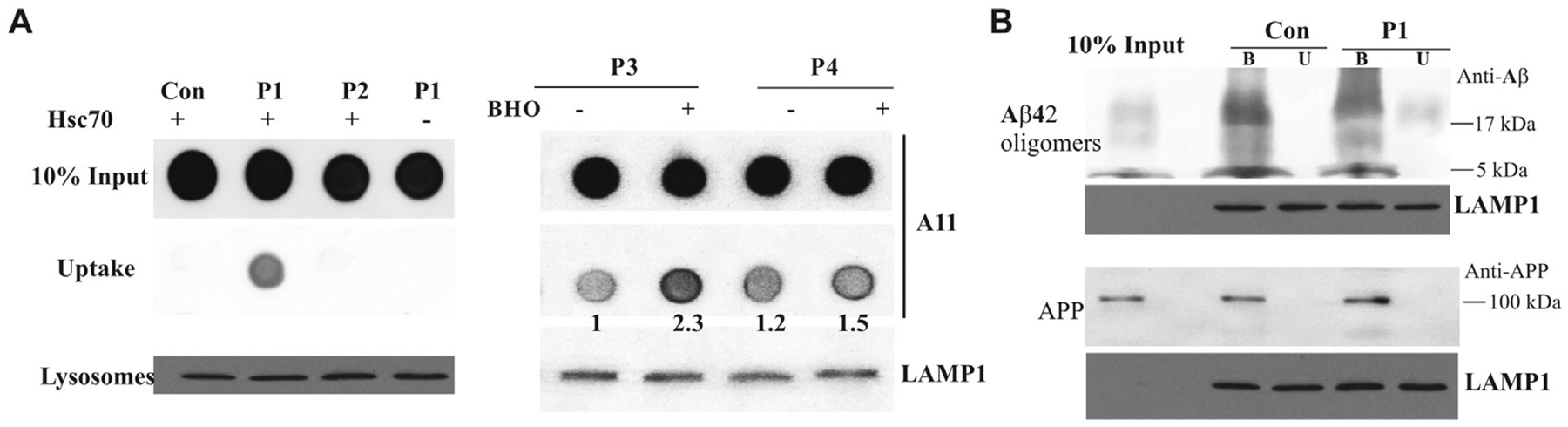
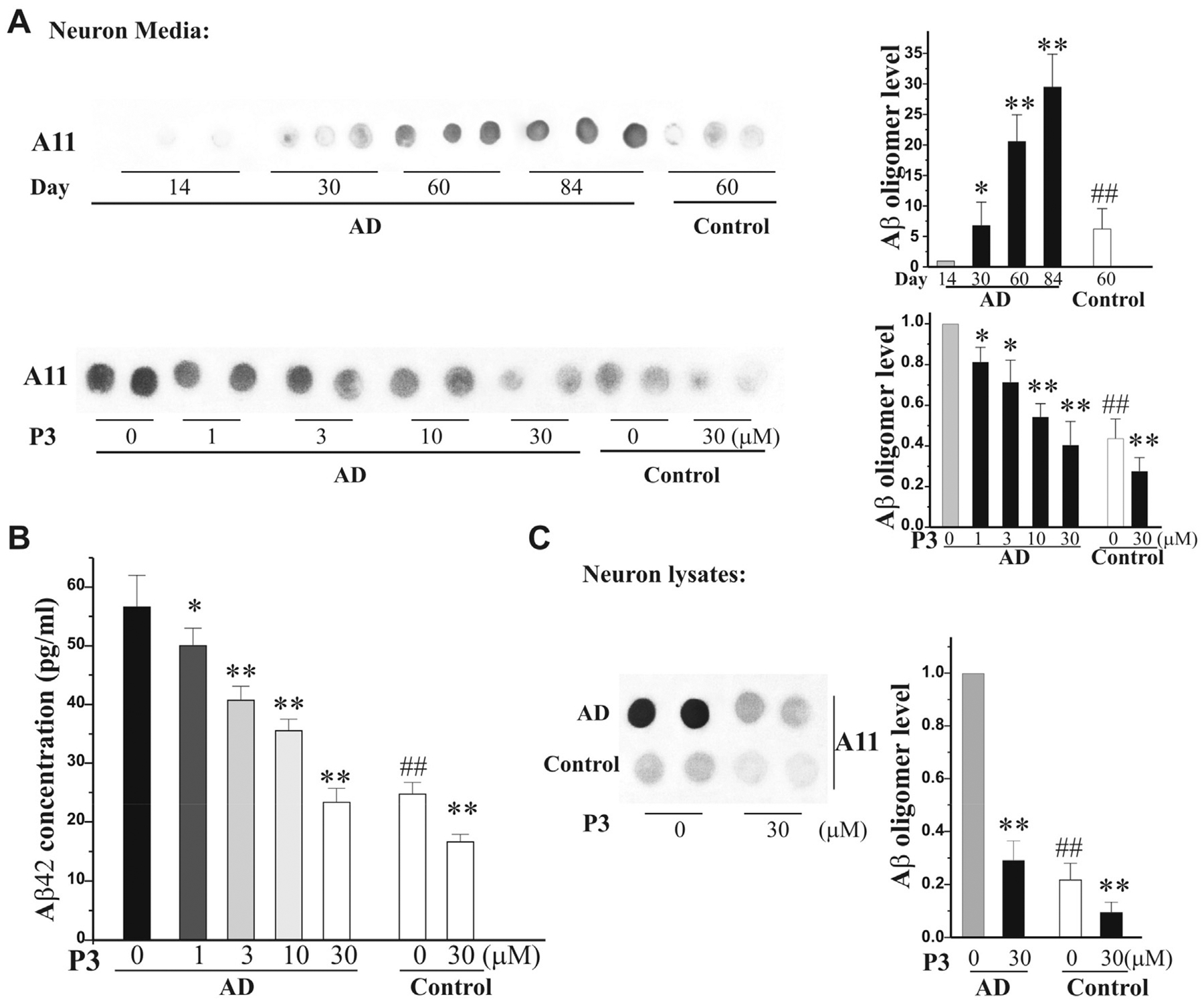
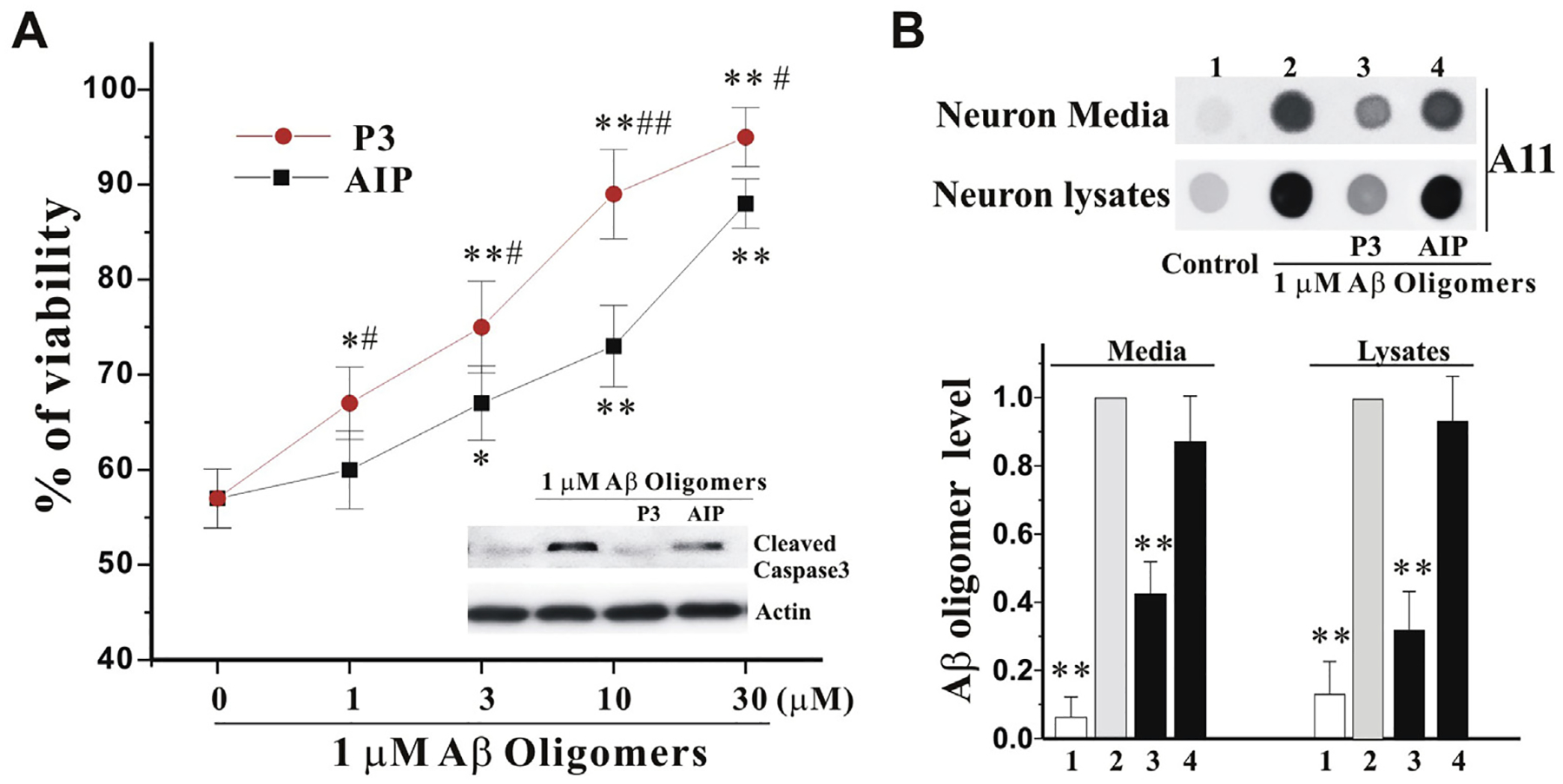
Amyloid β (Aβ) oligomers may be a real culprit in the pathogenesis of Alzheimer's disease (AD); therefore, the elimination of these toxic oligomers may be of great significance for AD therapy. Autophagy is the catabolic process by which lysosomes degrade cytosolic components, and heat shock cognate 70 kDa protein (Hsc70) binds to proteins with their KFERQ-like motifs [also known as chaperone-mediated autophagy (CMA) motifs] and carries them to lysosomes through CMA or late endosomes through endosomal microautophagy (eMI) for degradation. In this study, our strategy is to make the pathological Aβ become one selective and suitable substrate for CMA and eMI (termed as Hsc70-based autophagy) by tagging its oligomers with multiple CMA motifs. First, we design and synthesize Aβ oligomer binding peptides with three CMA motifs. Second, we determine that the peptide can help Aβ oligomers enter endosomes and lysosomes, which can be further enhanced by ketone. More importantly, we find that the peptide can dramatically reduce Aβ oligomers in induced pluripotent stem cell (iPSC) cortical neurons derived from AD patient fibroblasts and protect primary cultured cortical neurons against the Aβ oligomer-induced neurotoxicity. In conclusion, we demonstrate that the peptide targeting Hsc70-based autophagy can effectively eliminate Aβ oligomers and have superior neuroprotective activity.

Keywords: Alzheimer’s disease; Aβ oligomers; Chaperone-mediated autophagy; Hsc70.


Conflict of interest statement

Declaration of competing interest The authors declare that they have no known competing financial interests or personal relationships that could have appeared to influence the work reported in this paper.

Figures

Fig. 1.
Fig. 1.. Interacting peptide sequence to Aβ oligomers and proposed its mechanisms of action.
A. Three domains of the peptides: TAT, BD (Binding domain), and CTM (Top panel). The peptides with three readily or hard oxidized amino acids (OD, oxidized domain) (Bottom panel). B. Proposed mechanism of action of novel peptides: the novel peptides bind to Aβ oligomers, the peptide-oligomer complexes may be recognized by Hsc70, and Hsc70 brings Aβ oligomers into endosomes and lysosomes in cells for degradation. C. Determination of novel peptide with different formats of Ab: 0.5 μg of soluble Aβ42 monomers, oligomers, and fibrils was applied to a nitrocellulose membrane and probed with biotin-P1, anti-oligomer A11 (AHB0052, Invitrogen), and anti-Aβ antibody (Cat. 8243, Cell signaling). D. Novel peptide specifically pulls down Aβ42 oligomers: 5 μg of soluble Aβ42 monomers, oligomers, and fibrils were incubated with Biotin-P1 for 30 min and then pulled down with the Streptavidin magnetic beads. The pellets were for dot assay probed with the anti-Aβ antibody.
Fig. 2.
Fig. 2.. Peptides P1 and P3 bring Aβ oligomers into endosomes/lysosomes.
A. Aβ oligomers were prepared for the uptake assay and incubated with indicated novel peptides with/out Hsc70. The matrix of endosomes/lysosomes was collected for the dot blot assay with the antibody A11 (Left panel). Aβ oligomers were prepared for uptake assay and incubated with indicated novel peptides with/out BHO. The matrix of endosomes/lysosomes was collected for the dot blot assay with the antibody A11 (Right panel). B. Aβ oligomers and APP were prepared for purified lysosome binding and uptake assays with/out P1, and then collected for Western blot assay with anti-Aβ and Anti-APP antibodies. The letters B and U stand for binding and uptake assay, respectively. LAMP1 serves as controls for all uptake and binding assays.
Fig. 3.
Fig. 3.. Peptide P3 reduces Aβ oligomers in hiPSC neurons.
A. P3 reduces Aβ oligomers in the media of AD iPSC neurons: Aβ oligomers increase in the media of AD iPSC neurons following culture at day 14–84. At day 60, the AD iPSC neurons were treated with P3 (1–30 μM) and the control iPSC neurons were treated with P3 (30 μM). The conditioned media (10 μl = 2 μl × 5 per dot) were collected for dot blot assay with A11 antibody. Left panels showed the quantification of Aβ oligomers relative to indicated groups (n = 3 of three independent experiments). B. The media were collected for the Aβ42 ELISA assay. C. The neuron lysates (6 mg per dot) were collected at the above same condition in the presence of P3 at 30 μM for dot blot assay. Left panel showed the quantification of Aβ oligomers relative to indicated groups (n = 3 of three independent experiments). All data in A to C were expressed as means ± SEM. n = 3, *p < 0.05, **P < 0.01 vs its corresponding control (without P3); ##P < 0.01 vs the AD iPSC neuron group.
Fig. 4.
Fig. 4.. Peptide P3 protects neurons from neurotoxicity of Aβ oligomers.
A. Primary cultured cortical neurons were treated with 6-h pre-incubated Aβ oligomers (1 μM), in the presence of indicated increase concentrations of P3 and AIP for 42 h. The viability (%) was measured by WST1 and expressed as mean ± SEM, normalized to vehicle-treated neurons. N = 6, *p < 0.05, **P < 0.01 vs control, and #p < 0.05, ##p < 0.01 vs AIP at the same concentration. The neuron lysates were collected from the above samples in the presence of both P3 and AIP at 30 mM for Western blot with cleaved caspase 3 antibody (Inset). B. The neurons were treated as the above in the presence of both P3 and AIP at 30 μM for 42 h. The neuron media (6 μl = 2 μl × 3 per dot) and lysates (4 μg per dot) were collected for dot blot assay with anti-A11 antibody (Top panel). Bottom panel showed the quantification of Aβ oligomers relative to Aβ oligomers alone group (n = 3 of independent experiments). All values are means ± SEM (**P < 0.01 vs the Aβ oligomers alone group).

Similar articles

Cited by

References

    1. Zendehbad AS, Noroozian M, Shakiba A, Kargar A, Davoudkhani M, Validation of Iranian Smell Identification Test for screening of mild cognitive impairment and Alzheimer’s disease, Appl. Neuropsychol. Adult (2020) 1–6, 10.1080/23279095.2019.1710508. - DOI - PubMed
    1. Selkoe DJ, Hardy J, The amyloid hypothesis of Alzheimer’s disease at 25 years, EMBO Mol. Med 8 (2016) 595–608, 10.15252/emmm.201606210. - DOI - PMC - PubMed
    1. Luo J, Warmlander SK, Graslund A, Abrahams JP, Cross-interactions between the alzheimer disease amyloid-beta peptide and other amyloid proteins: a further aspect of the amyloid cascade hypothesis, J. Biol. Chem 291 (2016) 16485–16493, 10.1074/jbc.R116.714576. - DOI - PMC - PubMed
    1. Lasagna-Reeves CA, Castillo-Carranza DL, Sengupta U, Sarmiento J, Troncoso J, Jackson GR, Kayed R, Identification of oligomers at early stages of tau aggregation in Alzheimer’s disease, FASEB J 26 (2012) 1946–1959, 10.1096/fj.11-199851. - DOI - PMC - PubMed
    1. Spires-Jones TL, Hyman BT, The intersection of amyloid beta and tau at synapses in Alzheimer’s disease, Neuron 82 (2014) 756–771, 10.1016/j.neuron.2014.05.004. - DOI - PMC - PubMed

Publication types

MeSH terms

LinkOut - more resources