Review
. 2020 Apr 1:106:34-53.
doi: 10.1016/j.actbio.2020.02.010. Epub 2020 Feb 11.

Computational modeling of cardiac growth and remodeling in pressure overloaded hearts-Linking microstructure to organ phenotype

Affiliations
Review

Computational modeling of cardiac growth and remodeling in pressure overloaded hearts-Linking microstructure to organ phenotype

Justyna A Niestrawska et al. Acta Biomater. .

Abstract

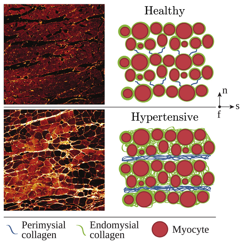
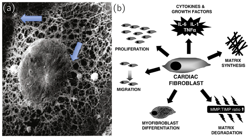
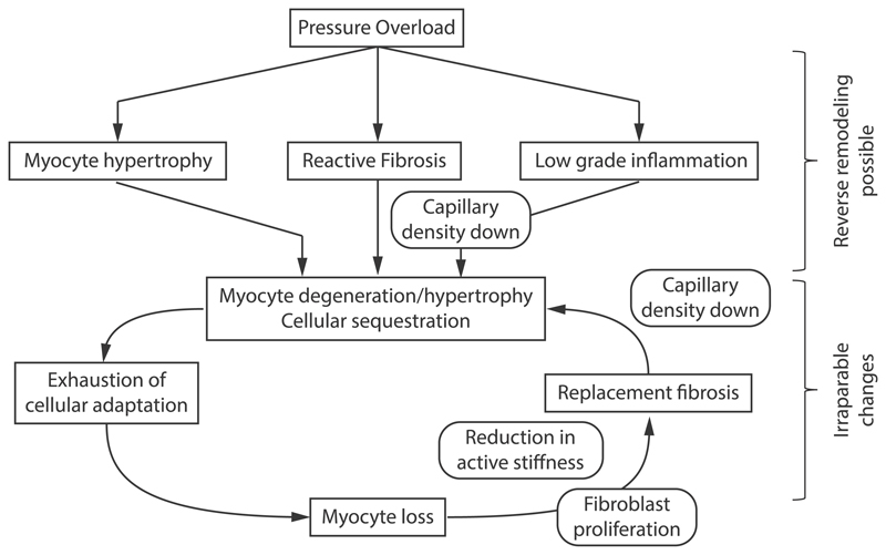
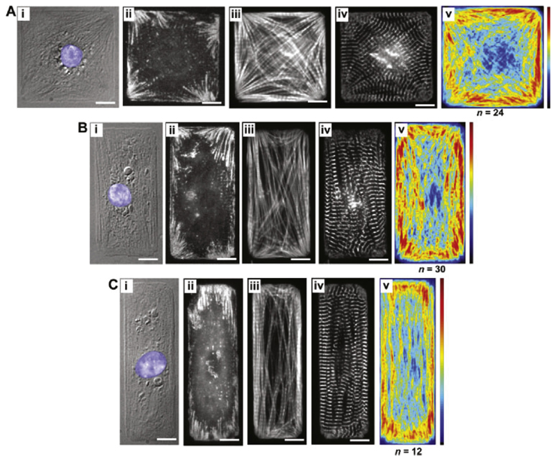
Cardiac growth and remodeling (G&R) refers to structural changes in myocardial tissue in response to chronic alterations in loading conditions. One such condition is pressure overload where elevated wall stresses stimulate the growth in cardiomyocyte thickness, associated with a phenotype of concentric hypertrophy at the organ scale, and promote fibrosis. The initial hypertrophic response can be considered adaptive and beneficial by favoring myocyte survival, but over time if pressure overload conditions persist, maladaptive mechanisms favoring cell death and fibrosis start to dominate, ultimately mediating the transition towards an overt heart failure phenotype. The underlying mechanisms linking biological factors at the myocyte level to biomechanical factors at the systemic and organ level remain poorly understood. Computational models of G&R show high promise as a unique framework for providing a quantitative link between myocardial stresses and strains at the organ scale to biological regulatory processes at the cellular level which govern the hypertrophic response. However, microstructurally motivated, rigorously validated computational models of G&R are still in their infancy. This article provides an overview of the current state-of-the-art of computational models to study cardiac G&R. The microstructure and mechanosensing/mechanotransduction within cells of the myocardium is discussed and quantitative data from previous experimental and clinical studies is summarized. We conclude with a discussion of major challenges and possible directions of future research that can advance the current state of cardiac G&R computational modeling. STATEMENT OF SIGNIFICANCE: The mechanistic links between organ-scale biomechanics and biological factors at the cellular size scale remain poorly understood as these are largely elusive to investigations using experimental methodology alone. Computational G&R models show high promise to establish quantitative links which allow more mechanistic insight into adaptation mechanisms and may be used as a tool for stratifying the state and predict the progression of disease in the clinic. This review provides a comprehensive overview of research in this domain including a summary of experimental data. Thus, this study may serve as a basis for the further development of more advanced G&R models which are suitable for making clinical predictions on disease progression or for testing hypotheses on pathogenic mechanisms using in-silico models.

Keywords: Computational modeling; Growth and remodeling; Hypertrophy; Pressure overload; Structural remodeling.


Conflict of interest statement

Declaration of Competing Interest The authors declare that the research was conducted in the absence of any commercial or financial relationships that could be construed as a potential conflict of interest.

Figures

Fig. 1
Fig. 1
In kinematic growth theory, a body is deformed due to growth and external loads in two time points τ and s. The total deformation gradient is decomposed into an inelastic growth part Fg and an elastic part Fe, leading to geometric compatibility and mechanical equilibrium. Adapted with permission from Cyron and Humphrey [25].
Fig. 2
Fig. 2
In constrained mixture models, a body is composed by n individual constituents, each consisting of multiple mass increments which were deposited with a prestretch Fpre(t) at different times. The elastic pre-stretch depends on the individual stress-free natural configuration of each constituent. All constituents undergo the same elastic deformation together, despite having been deposited with different pre-stretches at different times. Adapted with permission from Cyron and Humphrey [25].
Fig. 3
Fig. 3
(a) Schematic drawing of the arrangement of myocytes, reproduced with permission from Wang et al. [115]; (b) Schematic drawing of the structure of the myocyte, reproduced with permission from Kaplan [127].
Fig. 4
Fig. 4
(a) Scanning electron micrographs showing endomysial collagen surrounding myocyte lacunae (M) and a lacunae of a capillary (C) surrounded by the same collagen, scale bar 6.5 μm. (b) Schematic drawing of the distribution of endomysial collagen in a rabbit heart; a collagen weave (CW) enveloping myocytes (M) and capillaries (C) and collagen structs (CS) connecting single myocytes to each other. (a) and (b) modified with permission from Macchiarelli et al. [139]. (c) Scanning electron micrographs of struts of perimysial collagen anchored to the sarcolemma surface at the Z band plane (Z), scale bar 10 μm, and (d) a schematic drawing of this structure. S: sarcolemma, St: strut, IF: intermediate filament, M: mitochondrion, SSD: subsarcolemmal density, C: collagen. (c) and (d) modified with permission from Robinson et al. [147].
Fig. 5
Fig. 5
High resolution ex vivo confocal images of tissue blocks from a healthy rat heart and schematics showing the arrangement of the constituents (top) and high resolution ex vivo confocal images showing a rat heart after remodeling due to pressure overload and the corresponding schematic (bottom), adapted with permission from Wang et al. [115].
Fig. 6
Fig. 6
(a) A fibroblast with removed plasma showing the cytoskeletal network connecting an adhesion side on the plasma membrane with the connection to the nuclear membrane (arrows). Reproduced with permission from [175]. (b) A schematic depicting the various responses of a cardiac fibroblast to environmental stimuli, including differentiation into another phenotype, migration, contribution to ECM turnover, secretion of growth factors and matrix degradation. Reproduced with permission from [148].
Fig. 7
Fig. 7
A schematic showing the pathogenesis of pressure overload induced hypertrophy. Based on [3].
Fig. 8
Fig. 8
Bray et al. [135] printed ECM islands and placed myocytes on them to study the influence of the ECM on intracellular constituent alignment. The three cellular aspects are (A): 1:1, (B): 2:1 and (C): 3:1. (i) depicts a DIC image, (ii)–(iv) immunofluorescent stains for vinculin (revealing focal adhesions), F-actin (staining I-bands) and sarcomeric α-actin (revealing Z-bands). The average distribution of F-actin is shown in (v). Reproduced with permission from [135].
Fig. 9
Fig. 9
A schematic showing the interaction between thin and intermediate filaments within the titin/Z-disc complex with focal adhesion complexes, hence serving as mechanosensors. Titin has elastic sequences in the I-band, serving as springs saving elastic energy during diastole and relasing it to regain the initial sarcomere length at systole. At peak diastole the titin elastic segments uncoil and add their contribution to ventricular wall distensibility. Increased stretch of the titin elastic segments is sensed and activates downstream signals for cardiac remodeling. Adapted with permission from [168].

References

    1. Opie LH, Commerford PJ, Gersh BJ, Pfeffer MA. Controversies in ventricular remodelling. Lancet. 2006;367:356–367. doi: 10.1016/S0140-6736(06)68074-4. - DOI - PubMed
    1. Grossman W, Jones D, McLaurin LP. Wall stress and patterns of hypertrophy in the human left ventricle. J Clin Invest. 1975;56:56–64. doi: 10.1172/JCI108079. - DOI - PMC - PubMed
    1. Hein S, Arnon E, Kostin S, Schönburg M, Elsässer A, Polyakova V, Bauer EP, Klövekorn W-P, Schaper J. Progression from compensated hypertrophy to failure in the pressure-overloaded human heart. Circulation. 2003;107:984–991. doi: 10.1161/01.CIR.0000051865.66123.B7. - DOI - PubMed
    1. Krayenbuehl HP, Hess OM, Monrad ES, Schneider J, Mall G, Turina M. Left ventricular myocardial structure in aortic valve disease before, intermediate, and late after aortic valve replacement. Circulation. 1989;79:744–755. doi: 10.1161/01.CIR.79.4.744. - DOI - PubMed
    1. Esposito G, Rapacciuolo A, Prasad SVN, Takaoka H, Thomas SA, Koch WJ, Rockman HA. Genetic alterations that inhibit in vivo pressure-overload hypertrophy prevent cardiac dysfunction despite increased wall stress. Circulation. 2002;105:85–92. - PubMed

Publication types