. 2020 Sep;154(6):662-672.
doi: 10.1111/jnc.14985. Epub 2020 Mar 13.

Role of 1-Deoxysphingolipids in docetaxel neurotoxicity

Affiliations

Role of 1-Deoxysphingolipids in docetaxel neurotoxicity

Katrin A Becker et al. J Neurochem. 2020 Sep.

Abstract

A major dose-limiting side effect of docetaxel chemotherapy is peripheral neuropathy. Patients' symptoms include pain, numbness, tingling and burning sensations, and motor weakness in the extremities. The molecular mechanism is currently not understood, and there are no treatments available. Previously, we have shown an association between neuropathy symptoms of patients treated with paclitaxel and the plasma levels of neurotoxic sphingolipids, the 1-deoxysphingolipids (1-deoxySL) (Kramer et al, FASEB J, 2015). 1-DeoxySL are produced when the first enzyme of the sphingolipid biosynthetic pathway, serine palmitoyltransferase (SPT), uses L-alanine as a substrate instead of its canonical amino acid substrate, L-serine. In the current investigation, we tested whether 1-deoxySL accumulate in the nervous system following systemic docetaxel treatment in mice. In dorsal root ganglia (DRG), we observed that docetaxel (45 mg/kg cumulative dose) significantly elevated the levels of 1-deoxySL and L-serine-derived ceramides, but not sphingosine-1-phosphate (S1P). S1P is a bioactive sphingolipid and a ligand for specific G-protein-coupled receptors. In the sciatic nerve, docetaxel decreased 1-deoxySL and ceramides. Moreover, we show that in primary DRG cultures, 1-deoxysphingosine produced neurite swellings that could be reversed with S1P. Our results demonstrate that docetaxel chemotherapy up-regulates sphingolipid metabolism in sensory neurons, leading to the accumulation of neurotoxic 1-deoxySL. We suggest that the neurotoxic effects of 1-deoxySL on axons can be reversed with S1P.

Keywords: 1-Deoxysphingolipids; Ceramide; Docetaxel-induced peripheral neuropathy; Serine palmitoyltransferase; Sphingosine-1-phosphate.


Conflict of interest statement

Conflict of interest disclosure. The authors declare no conflicts of interest.

Figures

Figure 1.
Figure 1.. Sphingolipid pathway.
A/A schematic representation of the sphingolipid pathway at the level of sphingoid bases and ceramides. 1-DeoxySL are produced when serine palmitoyl transferase uses L-alanine as the amino acid substrate. 1-DeoxySL species (derived from L-alanine) do not have an OH group at position C1, as do the sphingolipid species derived from L-serine.
Figure 2.
Figure 2.. Schematic representation of the docetaxel treatment and tissue collections time line.
C57BL/6JOlaHsd female mice, three (2h and 6h tissue collection) or four (24h tissue collection) per group, were used in the experiment. The time line is not in scale.
Figure 3.
Figure 3.. 1-DeoxySL species upregulated in the DRG of mice treated with docetaxel.
Mice (C57BL/6OlaHsd) were intraperitoneally injected three times (with four-week intervals) with docetaxel (cumulative dose 45mg/kg) or vehicle. DRG were harvested 6h and 24h after the third injection. Lipids were extracted from the isolated DRG and subjected to targeted mass spectrometry analyses of 1-deoxySL. Each treatment group consisted of three (control and 6h) or four (24h) mice. The statistical analyses are presented in Suppl. Tables 1 and 2. A/ C18 1-deoxydihydroceramide; B/ C20 1-deoxydihydroceramide; C/ C22 1-deoxydihydroceramide; D/ C22 1-deoxyceramide; E/ C24 1-deoxyceramide; and F/ 1-deoxysphingosine. The box plots were generated in Sigma plot 14.0 (Systat Software in San Jose, CA). For the treatment groups of three samples each line represents the actual lipid measurement. For the treatment group with four samples, the whiskers represent the higher and the lower value of the lipid measurement.
Figure 4.
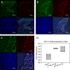
Figure 4.. SPTLC1 is upregulated in the DRG of mice treated with docetaxel.
Mice (C57BL/6OlaHsd) were intraperitoneally injected three times (with four-week intervals) with docetaxel (cumulative dose 45mg/kg). DRG were harvested 6h and 24h after the third injection of docetaxel or from the control group. Immunohistochemistry was performed on paraffin-fixed DRG slides and imaged by Nikon epifluorescence microscope. Nissl staining – green; SPTLC1 – red; DAPI- blue. A/ representative image of DRG isolated from control mouse; B/ DRG representative image of DRG isolated 6h after the 3rd injection from mouse treated with docetaxel; C/ representative image of DRG isolated 24h after the 3rd injection from mouse treated with docetaxel. D/ Nikon Elements analysis software was used to quantify the levels of fluorescence of Nissl and SPTLC1 labeling. All images were taken with the same microscope settings, and the same parameters of the software were used to analyze all of the images. DRG slides from three individual mice from each group were analyzed blindly as described in the material and methods. Technical repeats: three slides were used from each individual mouse/DRG with at least three regions of interest per slide. Statistical analyses (SAS Institute Inc., Cary, NC, USA): Ratio SPTLC1: Nissl, a Wilcoxon rank-sum test was performed to test for differences among Wilcoxon scores (ranks among the observations) between the control group and each treatment group. Significance was set at p<0.05. Control group compared to 6h after the 3rd docetaxel injection group p=0.0495. Control group compared to 24h after the 3rd docetaxel injection group p=0.0495.
Figure 5.
Figure 5.. 1-Deoxydihydrosphingolipid and 1-deoxySL species down-regulated in the sciatic nerve of mice treated with docetaxel.
Mice (C57BL/6OlaHsd) were intraperitoneally injected three times (with four-week intervals) with docetaxel (cumulative dose 45mg/kg). Sciatic nerves were harvested 6h (three mice) and 24h (four mice) after the third injection of docetaxel or from the control group (three mice). Lipids were extracted from the isolated nerves and subjected to targeted mass spectrometry analyses of 1-deoxySL. Each treatment group consisted of three or four mice. The statistical analyses are presented in Suppl. Table 8. A/ C18 1-deoxydihydroceramide; B/ C20 1-deoxydihydroceramide; C/ C22 1-deoxydihydroceramide; D/ 1-deoxysphinganine; E/ 1-deoxysphingosine. The box plots were generated in Sigma plot 14.0 (Systat Software in San Jose, CA). For the treatment groups of three samples each line represents the actual lipid measurement. For the treatment group with four samples, the whiskers represent the higher and the lower value of the lipid measurement.
Figure 6.
Figure 6.. Sphingosine-1-Phosphate (S1P) rescues 1-deoxysphingosine neurite swelling in primary DRG neurons.
The DRG neurons were isolated from C57BL/6 mice and cultured for 48h before labeled with SirActin (Cytoskeleton) for 6h and subsequently treated with Huzzah® conjugated lipids (Avanti Polar Lipids) for an additional 6h. Time-lapse imaging was perform on Nikon epifluorescence microscope. A – C/ Huzzah® control; D – F/ Huzzah®1-deoxysphingosine (500nM); white arrows in F show the neurite swellings; G – I/ Huzzah®1-deoxysphingosine (500nM) + Huzzah® S1P (500nM); J/ enlarged image from F; K/ neurite swellings quantification was performed blindly with Nikon Elements analysis software. The results are from three independent experiments; i.e. separate DRG neurons isolations. In each experiment, three individual neurons were imaged for each treatment and at least three neurite regions of interest were selected for each neuron. Statistical analyses; control vs. 1-deoxysphingosine (500nM) p<0.001; 1-deoxysphingosine vs. 1-deoxysphingosine (500nM) + S1P (500nM) p<0.001 (ANOVA, Sigma plot).

References

    1. Abram SE, Yi J, Fuchs A and Hogan QH (2006) Permeability of injured and intact peripheral nerves and dorsal root ganglia. Anesthesiology, 105, 146–153. - PubMed
    1. Alecu I, Othman A, Penno A, Saied EM, Arenz C, von Eckardstein A and Hornemann T (2017a) Cytotoxic 1-deoxysphingolipids are metabolized by a cytochrome P450-dependent pathway. J Lipid Res, 58, 60–71. - PMC - PubMed
    1. Alecu I, Tedeschi A, Behler N et al. (2017b) Localization of 1-deoxysphingolipids to mitochondria induces mitochondrial dysfunction. J Lipid Res, 58, 42–59. - PMC - PubMed
    1. Argyriou AA, Koltzenburg M, Polychronopoulos P, Papapetropoulos S and Kalofonos HP (2008) Peripheral nerve damage associated with administration of taxanes in patients with cancer. Crit Rev Oncol Hematol, 66, 218–228. - PubMed
    1. Bhatnagar B, Gilmore S, Goloubeva O, Pelser C, Medeiros M, Chumsri S, Tkaczuk K, Edelman M and Bao T (2014) Chemotherapy dose reduction due to chemotherapy induced peripheral neuropathy in breast cancer patients receiving chemotherapy in the neoadjuvant or adjuvant settings: a single-center experience. Springerplus, 3, 366. - PMC - PubMed

Publication types

MeSH terms