A 5 years follow-up for ischemic cardiac outcomes in patients with carotid artery calcification on panoramic radiographs confirmed by doppler ultrasonography in Turkish population
- PMID: 32058807
- PMCID: PMC7213528
- DOI: 10.1259/dmfr.20190440
A 5 years follow-up for ischemic cardiac outcomes in patients with carotid artery calcification on panoramic radiographs confirmed by doppler ultrasonography in Turkish population
Abstract
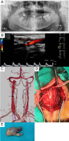
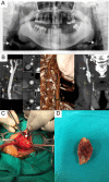
Objective: To evaluate the diagnostic accuracy of digital panoramic radiograph (DPR) for detection of carotid artery calcification (CAC) confirmed by Doppler Ultrasonography (DUSG) and to clarify the relationship between between CAC identified by DPR and cardiovascular events through a 5 year follow-up period.
Methods: Of 3600 consecutive patients examined, 158 patients presented with CAC as detected by DPR. The final study group was composed of 96 patients who had CAC confirmed by DUSG or CT angiogram. The control group was composed of 62 patients who has normal DUSG. The end point of the study was the occurrence of any cardiovascular event.
Results: 72 (75%) of the 96 patients with CAC confirmed by DUSG (16 patients had significant stenosis) had bilateral and 24 (25%) had unilateral CAS as detected by DUSG. There was a low agreement between the examination results with a κ value of 0.488 (p < 0.005) for calcification. Study data revealed that smoking, chronic obstructive pulmonary disease (COPD), diabetes mellitus (DM) and diastolic hypertension were significantly more common in patients with CAC than the control group (p < 0.05). During the follow-up period, 13 subjects had myocardial infarction and 1 subject died; in the control group, 1 patient died after MI and 1 patient died of a non-cardiac event.
Conclusion: Patients with CAC detectable by DPR concomitant with COPD, DM, smoking or diastolic hypertension are more likely to suffer from vascular events. Therefore, patients with detectable carotid plaque in DPR require referral to a cardiovascular surgery clinic for further investigations.
Keywords: Carotid artery calcification; Panoramic radiography; Ultrasonography; cardiovascular events.
Figures


References
-
- Lee J-S, Kim O-S, Chung H-J, Kim Y-J, Kweon S-S, Lee Y-H, et al. . The prevalence and correlation of carotid artery calcification on panoramic radiographs and peripheral arterial disease in a population from the Republic of Korea: the Dong-gu study. Dentomaxillofac Radiol 2013; 42: 29725099. doi: 10.1259/dmfr/29725099 - DOI - PMC - PubMed

