. 2020 Feb:52:102658.
doi: 10.1016/j.ebiom.2020.102658. Epub 2020 Feb 12.

Mboat7 down-regulation by hyper-insulinemia induces fat accumulation in hepatocytes

Affiliations

Mboat7 down-regulation by hyper-insulinemia induces fat accumulation in hepatocytes

Marica Meroni et al. EBioMedicine. 2020 Feb.

Abstract

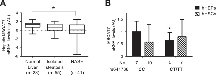
Background: Naturally occurring variation in Membrane-bound O-acyltransferase domain-containing 7 (MBOAT7), encoding for an enzyme involved in phosphatidylinositol acyl-chain remodelling, has been associated with fatty liver and hepatic disorders. Here, we examined the relationship between hepatic Mboat7 down-regulation and fat accumulation.

Methods: Hepatic MBOAT7 expression was surveyed in 119 obese individuals and in experimental models. MBOAT7 was acutely silenced by antisense oligonucleotides in C57Bl/6 mice, and by CRISPR/Cas9 in HepG2 hepatocytes.

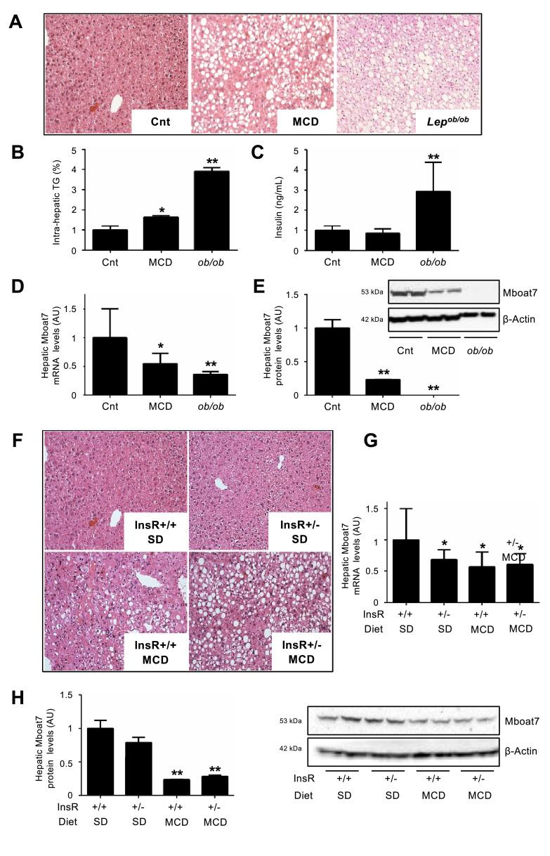
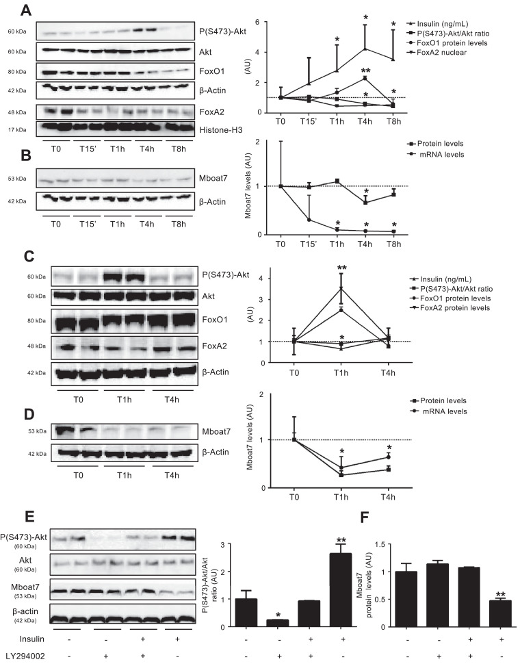
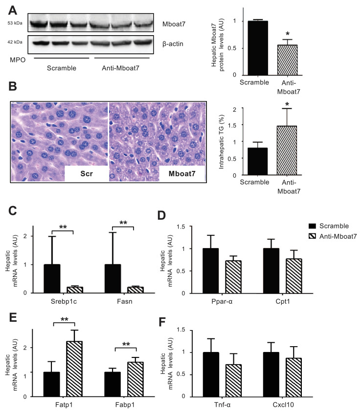
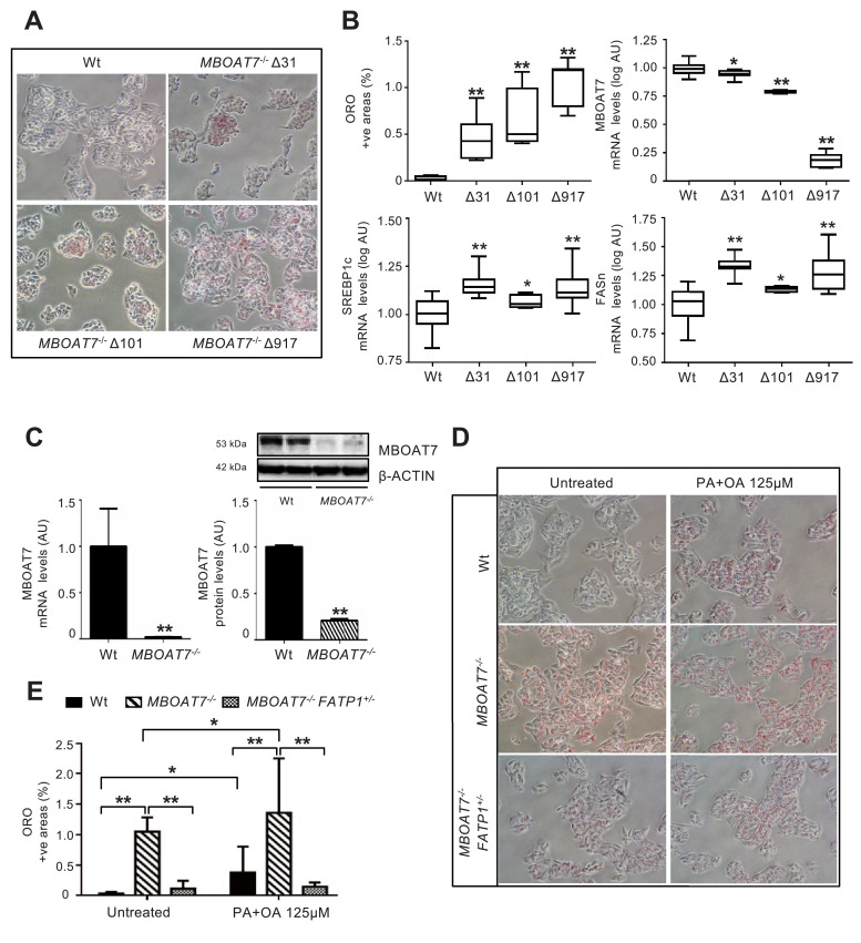
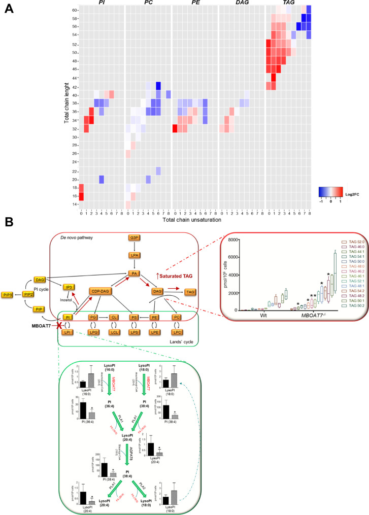
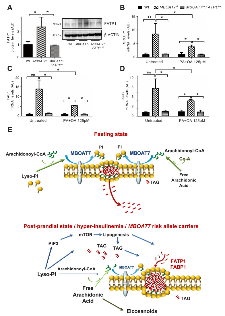
Findings: In obese individuals, hepatic MBOAT7 mRNA decreased from normal liver to steatohepatitis, independently of diabetes, inflammation and MBOAT7 genotype. Hepatic MBOAT7 levels were reduced in murine models of fatty liver, and by hyper-insulinemia. In wild-type mice, Mboat7 was down-regulated by refeeding and insulin, concomitantly with insulin signalling activation. Acute hepatic Mboat7 silencing promoted hepatic steatosis in vivo and enhanced expression of fatty acid transporter Fatp1. MBOAT7 deletion in hepatocytes reduced the incorporation of arachidonic acid into phosphatidylinositol, consistently with decreased enzymatic activity, determining the accumulation of saturated triglycerides, enhanced lipogenesis and FATP1 expression, while FATP1 deletion rescued the phenotype.

Interpretation: MBOAT7 down-regulation by hyper-insulinemia contributes to hepatic fat accumulation, impairing phosphatidylinositol remodelling and up-regulating FATP1.

Funding: LV was supported by MyFirst Grant AIRC n.16888, Ricerca Finalizzata Ministero della Salute RF-2016-02,364,358, Ricerca corrente Fondazione IRCCS Ca' Granda Ospedale Maggiore Policlinico; LV and AG received funding from the European Union Programme Horizon 2020 (No. 777,377) for the project LITMUS-"Liver Investigation: Testing Marker Utility in Steatohepatitis". MM was supported by Fondazione Italiana per lo Studio del Fegato (AISF) 'Mario Coppo' fellowship.

Keywords: LPIAT1; NAFLD; Nash; Nonalcoholic fatty liver disease; Phosphatidylinositol; Phospholipid; Steatohepatitis.


Figures

Fig 1
Fig. 1
Hepatic MBOAT7 expression decreases with liver damage severity and MBOAT7 rs641738 C>T affects MBOAT7 expression in human hepatocytes. Hepatic MBOAT7 mRNA levels were evaluated by qRT-PCR in 119 severely obese patients stratified by liver damage severity (A). Demographic, anthropometric and clinical characteristics of these patients are shown in Table S1. Boxes span from 25° to 75° percentile, while whiskers indicate the 10° and 90° percentile. Data is normalized to β-actin, log-transformed and expressed as fold increase (Arbitrary Units - AU). * adjusted p<0.05. MBOAT7 mRNA levels were evaluated by qRT-PCR in freshly isolated human hepatocytes (hHEPS) and activated human hepatic stellate cells (hHSCs, at third passage) stratified by the presence of MBOAT7 rs641738 T allele. The mRNA levels were normalized for β-actin and expressed as fold increase (Arbitrary Units - AU), as compared to CC genotype. (n = 17 hHEPs; n = 12 hHSCs) *p<0.05 compared to hHEPs or hHSCs carrying the protective CC genotype by two-tailed Student's t-tests (B).
Fig 2
Fig. 2
Hepatic Mboat7 expression is down-regulated in experimental models of NAFLD/NASH and hyper-insulinemia. Hematoxylin and Eosin (H&E) stain of hepatic specimens (A) and hepatic triglyceride (TG) content (B) from wild-type mice fed standard diet (referred to as Cnt) or Methionine choline deficient diet (MCD) and from Lepob/ob mice. Original magnification: 200X. Serum insulin was measured in mice fasted for 16 h (n = 10 per group) (C). Hepatic Mboat7 mRNA levels were evaluated by qRT-PCR, and mRNA levels were normalized for β-actin and are expressed as fold increase (Arbitrary Units - AU), as compared to Cnt (D). Western blot and quantification of Mboat7 protein levels in livers from Cnt, MCD and Lepob/ob mice, fasted 16 h (n = 10 per group) (E). * adjusted p<0.05, ** adjusted p<0.01 compared to Cnt. H&E stain of hepatic specimens from wild-type and InsR+/- mice fed standard diet (SD) or MCD (F). Original magnification: 200X. Hepatic Mboat7 mRNA levels were evaluated by qRT-PCR. mRNA levels were normalized for β-actin and expressed as fold increase (Arbitrary Units - AU), as compared to wild-type SD (InsR+/+ SD) (G). Western blot and quantification of Mboat7 protein levels in livers pooled from wild-type and InsR+/- MCD mice, fasted for 16 h (n = 10 per group) (H). * adjusted p<0.05; ** adjusted p<0.01 compared to InsR+/+ SD. All reactions were performed in duplicate in the same gel.
Fig 3
Fig. 3
Hepatic Mboat7 expression is modulated by nutritional state and insulin. Serum insulin (ng/mL) and Western blot and quantification of hepatic pSer473-Akt/Akt ratio, FoxO1, nuclear FoxA2 (A) and Mboat7 (B) protein levels evaluated in wild-type mice, fasted for 16 h (T0) or refed for 15 min (T15’), 1 h (T1h), 4 h (T4h) or 8 h (T8h) (n = 7 per group). FoxA2 nuclear fraction was normalized for Histone-H3 protein levels. Mboat7 mRNA levels were evaluated by qRT-PCR, normalized for β-actin and expressed as fold increase (Arbitrary Units - AU), as compared to T0. *p < 0.05, **p < 0.01 compared to T0. Western blot and quantification of hepatic pSer473-Akt/Akt ratio, FoxO1, FoxA2 (C) and Mboat7 (D) protein levels and serum insulin evaluated in wild-type mice, fasted for 16 h (T0) or 1 h (T1) and 4 h (T4) after insulin administration (0.6 U/Kg i.p, n = 7 per group). *p < 0.05; **p < 0.01 compared to T0. Western blot and quantification of pSer473-Akt/Akt ratio (E) and Mboat7 protein levels (F) in hepatocytes isolated from wild-type mice (n = 3) and treated with insulin (0.33 μM), LY294002 (50 μM) or a combination of both for 6 h; * adjusted p < 0.05, ** adjusted p < 0.01 compared to wild-type untreated cells. At least three independent experiments were conducted. For each condition, freshly protein lysates were pooled. All reactions were performed in duplicate in the same gel.
Fig 4
Fig. 4
In vivo silencing of hepatic Mboat7 induces TAG accumulation in hepatocellular lipid droplets Wild-type mice were treated with anti-Mboat7-MPO (12.5 mg/kg) or scramble-MPO (Scr) administered i.v. daily for 4 days (n = 6 per group) and fasted for 16 h before sacrifice. Western blot quantification of Mboat7 hepatic protein levels were normalized for β-actin; *p<0.05 compared to Scr, by two-tailed Student's t-tests. For each condition, freshly protein lysates were pooled. All reactions were performed in triplicate in the same gel (A). H&E staining and intrahepatic TAG content assessment in anti-Mboat7-MPO or scramble-MPO mice (B). Original magnification: 400X. Hepatic Srebp1 and Fasn (genes involved in de novo lipogenesis) (C), Ppar-α and Cpt1 (genes involved in β-oxidation) (D), Fatp1 and Fabp1 (fatty acids transporters) (E), Tnf-α and Cxcl10 (inflammatory markers) (F) expression was evaluated by qRT-PCR; mRNA levels were normalized to β-actin. Data are expressed as fold increase compared to Scr mice (n = 6 mice per group; *p<0.05, **p<0.01 by two-tailed Student's t-tests.
Fig 5
Fig. 5
MBOAT7 deletion increases spontaneous fat accumulation in HepG2 hepatoma cells. Spontaneous fat accumulation in untreated MBOAT7−/− cells carrying Δ31, Δ101 and Δ917 was evaluated by ORO staining (200X magnification) and quantified by ImageJ software in 10 random non-overlapping micrographs for each experimental condition, by calculating the percentage of pixels above the threshold value with respect to the total pixels per area. At least three independent experiments were conducted (A). Impact of MBOAT7 deletions on hepatic fat, and MBOAT7, SREBP1c, FASn mRNA, evaluated by qRT-PCR then normalized for β-actin (Arbitrary Units - AU). Boxes span from 25° to 75° percentile, while whiskers indicate the 10° and 90° percentile (B). * adjusted p<0.05, ** adjusted p<0.01 compared to wild-type (Wt) cells. The Δ917 MBOAT7 deletion abrogates MBOAT7 expression, resulting in increased spontaneous fat accumulation, which was maintained after supplementation with palmitic and oleic acid (PA+OA 125 μM; ratio 1:2). mRNA levels were evaluated by qRT-PCR and protein levels by Western blot and then normalized for β-actin. For each condition, freshly protein lysates were pooled. All reactions were performed in duplicate in the same gel. **p<0.01 by two-tailed Student's t-tests (C). Lipid accumulation was evaluated by ORO staining (200X magnification) and quantified by ImageJ software in 10 random non-overlapping micrographs for each experimental condition, by calculating the percentage of pixels above the threshold value with respect to the total pixels per area. At least three independent experiments were conducted. * adjusted p<0.05, ** adjusted p<0.01 (D-E).
Fig 6
Fig. 6
Impact of MBOAT7 deletion on intracellular fat composition. A)Heatmaps, which were generated calculating log2 fold change ratio between MBOAT7−/−/wt absolute quantification. Red shading indicates the induction and blue shading indicates repression relative to the average levels of each lipid species across all samples. PI: phosphatidyl-inositol, PC: phosphatidyl-choline, PE: phosphatidyl-ethanolamine, DAG: diacylglycerol and TAG: triacylglycerols. B)Hypothetical model of the impact of reduced MBOAT7 activity on intracellular phospholipid metabolism in hepatocytes. The consequences of MBOAT7 downregulation are shown by the red cross and arrows. When MBOAT7 is not functional, LysoPI (20:4) cannot be generated from LysoPI (16:0) or LysoPI (18:0), which therefore accumulate, together with their precursors PI (36:4) and PI (38:4). Accumulating saturated-PIs are then shunted to the synthesis of TAGs. CDP: cytidine-diphosphate, CL: cardiolipin, DAG: diacylglycerol, TAG: triacylglycerols, G3P: glyceraldehyde-3-phosphate, LPA: lyso-phosphatidic acid, PA: phosphatidic acid, PC: phosphatidyl-choline, PE: phosphatidyl-ethanolamine, PG: phosphatidyl-glycerol, PI: phosphatidyl-inositol, PIP: phosphatidyl-inositol-phosphate, PS: phosphatidyl-serine (For interpretation of the references to color in this figure legend, the reader is referred to the web version of this article.).
Fig 7
Fig. 7
Fat accumulation in MBOAT7−/− hepatocytes requires FATP1. FATP1 protein levels were evaluated in lysates from wild-type (Wt), MBOAT7−/− and MBOAT7−/−/FATP1+/- cells by Western blot and then normalized for β-actin. For each condition, freshly protein lysates were pooled. All reactions were performed in duplicate in the same gel (A). * adjusted p<0.05 compared to Wt or MBOAT7−/− cells. In Wt, MBOAT7−/− and MBOAT7−/−/FATP1+/- cells SREBP1 (B), FASn (C) and ACC (D) gene expressions were evaluated by qRT-PCR and normalized for β-actin. * adjusted p<0.05; compared to Wt or MBOAT7−/− cells. Hypothetical mechanism(s) linking down-regulation of MBOAT7 in hepatocytes with hepatic fat accumulation (E). During fasting, MBOAT7 localizes to the endoplasmic reticulum and lipid droplets, where it conjugates arachidonoyl-CoA to the second acyl-chain of Lyso-PI allowing the incorporation into cell membranes. During hyperinsulinemia and in carriers of the rs641738 risk variant, MBOAT7 is reduced, increasing the concentration of saturated PI, which accumulate and are diverted to TAG synthesis. This process requires up-regulation of the fatty acid transporter FATP1, and in vitro is associated with increased de novo lipogenesis.

References

    1. Marchesini G., Brizi M., Bianchi G., Tomassetti S., Bugianesi E., Lenzi M. Nonalcoholic fatty liver disease: a feature of the metabolic syndrome. Diabetes. 2001;50(8):1844–1850. - PubMed
    1. Marchesini G., Brizi M., Morselli-Labate A.M., Bianchi G., Bugianesi E., McCullough A.J. Association of nonalcoholic fatty liver disease with insulin resistance. Am J Med. 1999;107(5):450–455. - PubMed
    1. Marra F., Gastaldelli A., Svegliati Baroni G., Tell G., Tiribelli C. Molecular basis and mechanisms of progression of non-alcoholic steatohepatitis. Trends Mol Med. 2008;14(2):72–81. - PubMed
    1. Dongiovanni P., Valenti L. Genetics of nonalcoholic fatty liver disease. Metab Clin Exp. 2016;65(8):1026–1037. - PubMed
    1. Dongiovanni P., Meroni M. miRNA signature in NAFLD: a turning point for a non-invasive diagnosis. 2018;19(12) - PMC - PubMed

MeSH terms