. 2020 Feb 21;23(2):100841.
doi: 10.1016/j.isci.2020.100841. Epub 2020 Jan 16.

Principles of Cell Circuits for Tissue Repair and Fibrosis

Affiliations

Principles of Cell Circuits for Tissue Repair and Fibrosis

Miri Adler et al. iScience. .

Abstract

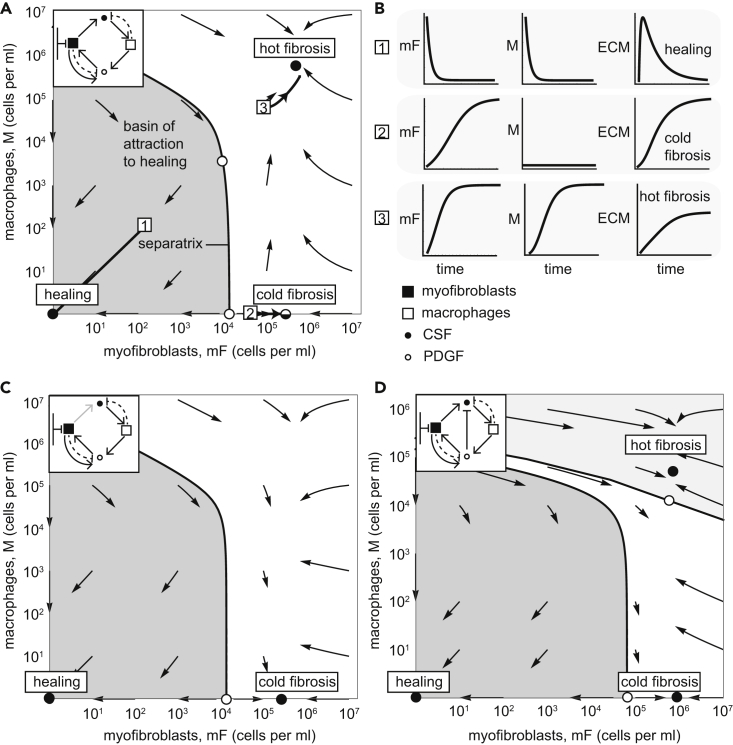
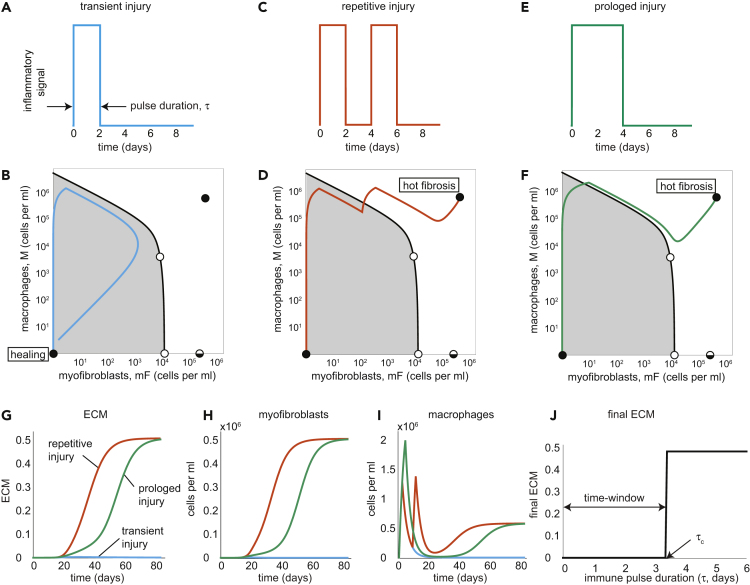
Tissue repair is a protective response after injury, but repetitive or prolonged injury can lead to fibrosis, a pathological state of excessive scarring. To pinpoint the dynamic mechanisms underlying fibrosis, it is important to understand the principles of the cell circuits that carry out tissue repair. In this study, we establish a cell-circuit framework for the myofibroblast-macrophage circuit in wound healing, including the accumulation of scar-forming extracellular matrix. We find that fibrosis results from multistability between three outcomes, which we term "hot fibrosis" characterized by many macrophages, "cold fibrosis" lacking macrophages, and normal wound healing. This framework clarifies several unexplained phenomena including the paradoxical effect of macrophage depletion, the limited time-window in which removing inflammation leads to healing, and why scar maturation takes months. We define key parameters that control the transition from healing to fibrosis, which may serve as potential targets for therapeutic reduction of fibrosis.

Keywords: In Silico Biology; Systems Biology; Tissue Engineering.


Conflict of interest statement

Declaration of Interest The authors declare no competing interests.

Figures

None
Graphical abstract
Figure 1
Figure 1
Overview of Cell-Cell Interactions Following Tissue Injury (A) Schematic of wound healing and scar formation. (B) If injury is transient, it leads to brief inflammation, and no fibrosis occurs. However, if injury is persistent and inflammation exceeds a critical time window, fibrosis is usually inevitable. (C) Circuit in which myofibroblasts (mF) and macrophages (M) secrete growth factors (GFs) for each other; mFs show an autocrine loop and are limited by a carrying capacity. Both cell types remove the GFs by endocytosis (dashed arrows). (D) ECM is produced by mF and degraded by proteases mainly secreted by M. Proteases are inhibited by factors secreted by mF and M.
Figure 2
Figure 2
The Myofibroblast-Macrophage Circuit Shows Multi-stability between a Healing State and Two Fibrosis States (A) Phase portrait of the circuit with reference parameters, in which arrows show the flow rate of change of cell numbers. Stable fixed points (black dots) and unstable fixed points (white dots) are shown (the semi-stable cold fibrosis state as split black and white), as is the separatrix (black line) that marks the boundary between the basin of attraction of the healing (gray region) and fibrosis states (the parameter values that are used are listed in Table 1). (B) Temporal trajectories of cells and ECM starting from the initial points 1–3 (white squares in panel B). (C) Phase portrait of a circuit with a 100-fold lower CSF secretion rate than in panel A stabilizes the cold-fibrosis state. There is no hot fibrosis state. (D) Myofibroblast-macrophage phase portrait in a circuit in which PDGF downregulates CSF expression in myofibroblast shows all three stable states. Note there are two separatrix curves, dividing the phase portrait into three basins of attraction. The middle basin (white region) flows to the cold-fibrosis state. Here we used the parameter values: α1=0.2moleculescellmin, α2=30moleculescellmin, β3=8moleculescellmin, β1=2moleculescellmin, λ1=λ2=21day. We used the values listed in Table 1 for the remaining model parameters. See also Figure S2.
Figure 3
Figure 3
The mF-M Circuit Shows Healing Versus Fibrosis Depending on Duration and Recurrence of Inflammation (A–I) (A) For a brief pulse of inflammation (2 days), the rise of mF and M is transient, leading to a healing trajectory in phase space that returns to the healing state (in light blue) (B). In contrast, two successive two-day long inflammatory pulses (C) or a prolonged pulse (4 days) (E) lead to a trajectory to the hot fibrosis state with persistent mF and M populations (in orange and green, respectively) (D and F). Note that the separatrix applies to the equations without the external inflammation input, and so the separatrix can be crossed during the input pulse(s). The dynamics of ECM (G), mFs (H), and Ms (I) are plotted in response to transient (light blue lines), repetitive (orange lines), and prolonged (green lines) injuries. (J) Final ECM as a function of inflammation pulse duration shows a critical time-window of about three days to stop inflammation. See also Figures S1, S3, and S4.
Figure 4
Figure 4
The mF-M Circuit Explains the Timescale of Months for Scar Maturation and the Paradoxical Effect of Macrophage Depletion on Fibrosis (A) ECM accumulation in response to a four-day immune pulse. Maturation time, tm, is defined as the time to reach half maximal ECM accumulation. (B–F) (B) ECM maturation time is on the order of months for fibrosis and weeks for healing. The slow timescale of weeks to months is due to a dynamical barrier due to an unstable fixed point (upper white circle) (C and D), akin to a ball slowing down at the top of a hill (E and F). In (C and D) blue dots indicate trajectory values at intervals of one day, so that slow dynamics correspond to dense blue dots. (G) Depleting macrophages when myofibroblasts are below or above the unstable point (lower white circle, labeled mFu) leads to healing or fibrosis, respectively.
Figure 5
Figure 5
Fibrosis Can Be Prevented or Reversed by Changing Several Circuit Parameters (A) Critical time window for inflammation that results in healing as a function of fold-change in circuit parameters. The window can be lengthened by decreasing autocrine secretion, increasing PDGF endocytosis, or decreasing the ratio of mF proliferation to removal. (B) Increasing PDGF endocytosis rate by 150% eliminates the cold fibrosis state, enlarging the basin of attraction to the healing state (gray region) and allowing a four-day immune pulse to resolve in healing. (C) Dynamics of myofibroblasts (black) and macrophages (blue) following a four-day immune pulse that lead to fibrosis without treatment (dashed curves). A 14-day treatment in which the PDGF endocytosis rate is increased by 150% that is given 20 days after the injury leads to healing. (D) The same 14-day treatment is shown in a phase portrait of the cells. The timing of the treatment is given when the cells are below the separatrix with the altered parameters (in red), and the duration of the treatment lasts until the cells cross the original separatrix (in black). (E) The effect of changing the PDGF endocytosis rate 20 days following a four-day immune pulse is shown for different values of duration and dose of the treatment. (F) Reversing a mature scar following a four-day immune pulse by increasing PDGF endocytosis rate by 300% for 40 days. (G) Reversing the same mature scar by a 25-day treatment of 300% larger PDGF endocytosis rate following macrophage depletion.
Figure 6
Figure 6
Overview of the Present Circuit Approach to understanding Healing and Fibrosis (A) The mF-M circuit shows a stable healing state and two fibrosis states. Outcome depends on the duration and persistence of inflammation pulses caused by an injury, which can remain in the basin of attraction for healing, or cross the separatrix into the basin of attraction for fibrosis. (B) The present analysis suggests targets for prevention and reversal of fibrosis, by eliminating the cold fibrosis fixed point and enlarging the basin of attraction to the healing state. See also Figure S5.

References

    1. Abergel R.P., Pizzurro D., Meeker C.A., Lask G., Matsuoka L.Y., Minor R.R., Chu M.-L., Uitto J. Biochemical composition of the connective tissue in keloids and analysis of collagen metabolism in keloid fibroblast cultures. J. Invest. Dermatol. 1985;84:384–390. - PubMed
    1. Adler M., Mayo A., Zhou X., Franklin R.A., Jacox J.B., Medzhitov R., Alon U. Endocytosis as a stabilizing mechanism for tissue homeostasis. Proc. Natl. Acad. Sci. U S A. 2018;115:E1926–E1935. - PMC - PubMed
    1. Adler M., Kohanim Y.K., Tendler A., Mayo A., Alon U. Continuum of gene-expression profiles provides spatial division of labor within a differentiated cell type. Cell Syst. 2019;8:43–52. - PubMed
    1. Alon, U., 2019. In: An Introduction to Systems Biology: Design Principles of Biological Circuits. CRC Press.
    1. Arno A.I., Gauglitz G.G., Barret J.P., Jeschke M.G. Up-to-date approach to manage keloids and hypertrophic scars: a useful guide. Burns. 2014;40:1255–1266. - PMC - PubMed

LinkOut - more resources