Clinical Trial
. 2020 Feb 14;15(2):e0228948.
doi: 10.1371/journal.pone.0228948. eCollection 2020.

Extracellular vesicles isolated from patients undergoing remote ischemic preconditioning decrease hypoxia-evoked apoptosis of cardiomyoblasts after isoflurane but not propofol exposure

Affiliations
Clinical Trial

Extracellular vesicles isolated from patients undergoing remote ischemic preconditioning decrease hypoxia-evoked apoptosis of cardiomyoblasts after isoflurane but not propofol exposure

Frederik Abel et al. PLoS One. .

Abstract

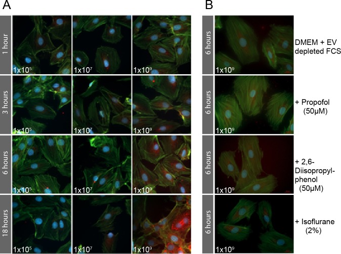
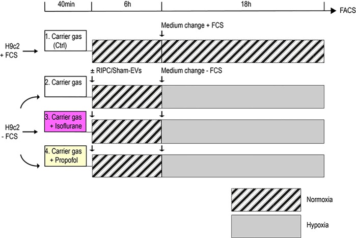
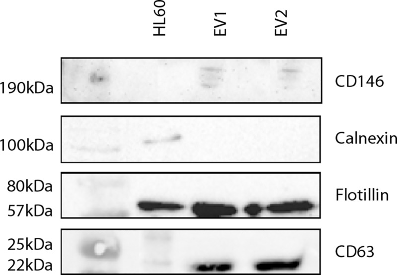
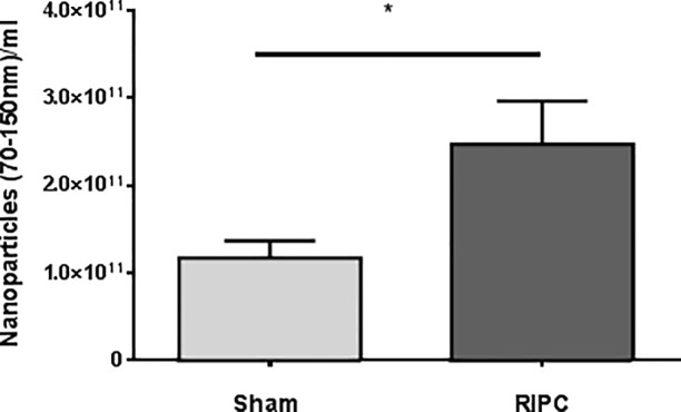
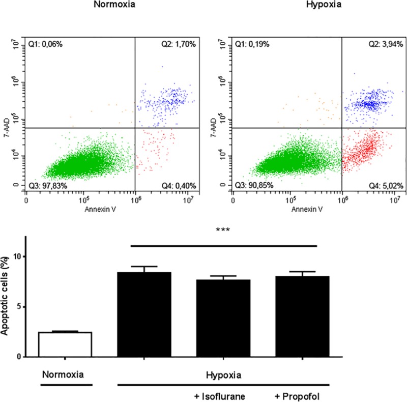
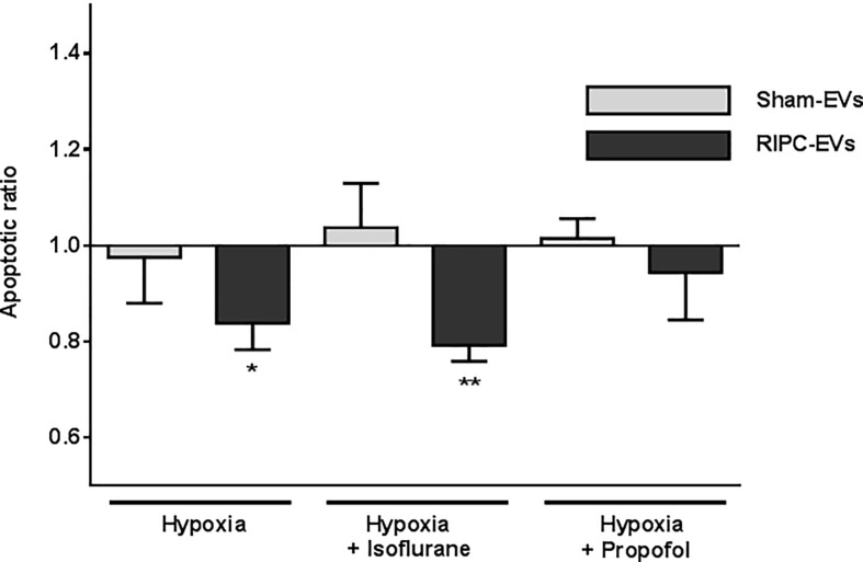
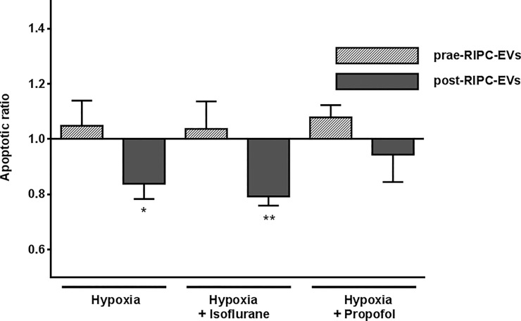
Remote ischemic preconditioning (RIPC) can evoke cardioprotection following ischemia/reperfusion and this may depend on the anesthetic used. We tested whether 1) extracellular vesicles (EVs) isolated from humans undergoing RIPC protect cardiomyoblasts against hypoxia-induced apoptosis and 2) this effect is altered by cardiomyoblast exposure to isoflurane or propofol. EVs were isolated before and 60 min after RIPC or Sham from ten patients undergoing coronary artery bypass graft surgery with isoflurane anesthesia and quantified by Nanoparticle Tracking Analysis. Following EV-treatment for 6 hours under exposure of isoflurane or propofol, rat H9c2 cardiomyoblasts were cultured for 18 hours in normoxic or hypoxic atmospheres. Apoptosis was detected by flow cytometry. Serum nanoparticle concentrations in patients had increased sixty minutes after RIPC compared to Sham (2.5x10¹¹±4.9x10¹⁰ nanoparticles/ml; Sham: 1.2x10¹¹±2.0x10¹⁰; p = 0.04). Hypoxia increased apoptosis of H9c2 cells (hypoxia: 8.4%±0.6; normoxia: 2.5%±0.1; p<0.0001). RIPC-EVs decreased H9c2 cell apoptosis compared to control (apoptotic ratio: 0.83; p = 0.0429) while Sham-EVs showed no protection (apoptotic ratio: 0.97). Prior isoflurane exposure in vitro even increased protection (RIPC-EVs/control, apoptotic ratio: 0.79; p = 0.0035; Sham-EVs/control, apoptotic ratio:1.04) while propofol (50μM) abrogated protection by RIPC-EVs (RIPC-EVs/control, Apoptotic ratio: 1.01; Sham-EVs/control, apoptotic ratio: 0.94; p = 0.602). Thus, EVs isolated from patients undergoing RIPC under isoflurane anesthesia protect H9c2 cardiomyoblasts against hypoxia-evoked apoptosis and this effect is abrogated by propofol. This supports a role of human RIPC-generated EVs in cardioprotection and underlines propofol as a possible confounder in RIPC-signaling mediated by EVs.


Conflict of interest statement

The authors have declared that no competing interests exist.

Figures

Fig 1
Fig 1
A. Intracellular uptake of BODIPY TR ceramide labeled EVs (red) into H9c2 cells. Association of EV concentration and incubation time in H9c2 cells as assessed by fluorescence microscope. Strongest fluorescence signals of intracellular EVs (red dots) were seen at an EV concentration of 1x10⁹ nanoparticles/ml with incubation periods of 1, 3, 6, and 18 hours with increased signaling intensity over time. Lesser concentrations of 1x10⁵ and 1x10⁷ part/ml did not evoke a detectable increase in fluorescence after any incubation time. Accordingly, a combination of 1x10⁹ nanoparticles/ml and 6 hours incubation time was used for further experiments to allow for adequate cellular EV uptake while avoiding potential oversaturation. B. Uptake of labeled EVs into H9c2 cells cultured with EV-depleted FCS Taking advantage of prior results, the experiment was repeated using the optimum incubation time and concentration for EV-uptake with DMEM and EV-depleted FCS (DMEM + FCS–EV) including four different setups: DMEM + FCS–EV, propofol soya emulsion in DMEM + FCS–EV, pure 2,6-diisopropylphenol in DMEM + FCS–EV, and isoflurane. As expected, the overall signal of labeled EVs in the cells was lower using EV-depleted FCS in compared to the first set of experiments with EV containing FCS. The cells show a similar EV-uptake in all conditions.
Fig 2
Fig 2. Experimental setup of H9c2 cells exposed to normoxia/hypoxia.
Each line represents a different condition. Cells were transferred to 6-well plates and incubated for 24 hours in standard DMEM + 25mM Hepes. The medium was then changed to fresh DMEM + 25mM Hepes ± FCS for each condition. All plates were perfused with normoxic gas (21% O2, 5% CO2), whereas one plate was additionally exposed to isoflurane 2% (magenta filled box) and one plate to 50μM propofol (yellow filled box). After 40 minutes EVs were added at a concentration of 1x10⁹ nanoparticles/ml. Normoxia treated cells did not receive EVs. After incubation for 6 hours, the medium was renewed again to DMEM + FCS in cells under normoxic conditions or to DMEM—FCS in cells undergoing hypoxia. H9c2 cells were then cultured in a normoxic or hypoxic atmosphere (1% O2, 5% CO2) for 18 hours. Apoptosis under all experimental conditions was measured by flow cytometry as the endpoint. The hatched boxes illustrate normoxic periods and the grey filled boxes hypoxic periods.
Fig 3
Fig 3. Western blots of EVs isolated from humans undergoing RIPC.
Presence of the cytosolic protein flotillin 1, EV marker CD63, endothelial cell marker CD146, and absence of calnexin was analyzed in two representative EV samples extracted of CABG-patients 60 min post-RIPC. HL60 cell lysate was used as a positive control.
Fig 4
Fig 4. Arterial nanoparticle concentration of patients after RIPC or Sham interventions as analyzed by Nanoparticle Tracking Analysis (NTA).
Total serum EV-concentration between 70-150nm/ml obtained from patients 60 minutes after RIPC or Sham treatment. Values are means ± SEM; n = 5 (*p<0.05).
Fig 5
Fig 5. A effect of normoxia/hypoxia exposure on apoptosis rate of H9c2 cells.
Representative flow cytometric images after 18 hours of normoxia or hypoxia without incubation with EVs. Apoptosis was detected by Annexin V/7-AAD staining. b Quantitative analysis of apoptotic cells after normoxia/hypoxia and isoflurane or propofol exposure. Apoptosis was markedly increased by hypoxia, whereas isoflurane or propofol had no significant additional beneficial effect. Results are expressed as means ± SEM; n = 10 (***p<0.0001).
Fig 6
Fig 6. Influence of RIPC-EV or Sham-EV fractions on apoptosis rate in H9c2 cells.
EVs were added to H9c2 cells at a concentration of 1x10⁹ nanoparticles/ml and incubated for 6 hours before a change of medium and for the following 18 hours of hypoxia. Apoptotic ratio of cells after hypoxia ± isoflurane or ± propofol plus EVs are presented compared to cells without EV incubation. Values are means ± SEM; n = 5 (*p<0.05, **p<0.01).
Fig 7
Fig 7. Impact of EV fractions on cell apoptosis isolated before RIPC in contrast to EVs isolated after RIPC.
Patient-derived EVs extracted before the RIPC maneuver (prae-RIPC-EVs) had no effect on apoptosis of H9c2 cells compared to cells incubated without EVs. In contrast to EV fractions isolated before the RIPC maneuver, post-RIPC EV fractions improved the apoptotic ratio of H9c2 cells after both hypoxia and hypoxia plus isoflurane. Data are presented as apoptotic ratio of cells with EV-coincubation compared to apoptosis without EV-coincubation. Values are means ± SEM; n = 5 (*p<0.05 **p<0.01).

References

    1. Heusch G. Cardioprotection: chances and challenges of its translation to the clinic. Lancet. 2013;381(9861):166–75. 10.1016/S0140-6736(12)60916-7 - DOI - PubMed
    1. Thielmann M, Kottenberg E, Kleinbongard P, Wendt D, Gedik N, Pasa S, et al. Cardioprotective and prognostic effects of remote ischaemic preconditioning in patients undergoing coronary artery bypass surgery: a single-centre randomised, double-blind, controlled trial. Lancet. 2013;382(9892):597–604. 10.1016/S0140-6736(13)61450-6 - DOI - PubMed
    1. Kleinbongard P, Peters J, Jakob H, Heusch G, Thielmann M. Persistent Survival Benefit From Remote Ischemic Pre-Conditioning in Patients Undergoing Coronary Artery Bypass Surgery. J Am Coll Cardiol. 2018;71(2):252–4. 10.1016/j.jacc.2017.10.083 - DOI - PubMed
    1. Pickard JM, Botker HE, Crimi G, Davidson B, Davidson SM, Dutka D, et al. Remote ischemic conditioning: from experimental observation to clinical application: report from the 8th Biennial Hatter Cardiovascular Institute Workshop. Basic Res Cardiol. 2015;110(1):453 10.1007/s00395-014-0453-6 - DOI - PMC - PubMed
    1. Hausenloy DJ, Candilio L, Evans R, Ariti C, Jenkins DP, Kolvekar S, et al. Remote Ischemic Preconditioning and Outcomes of Cardiac Surgery. N Engl J Med. 2015;373(15):1408–17. 10.1056/NEJMoa1413534 - DOI - PubMed

Publication types