. 2020 Feb 12;21(4):1229.
doi: 10.3390/ijms21041229.

Salicylic Acid Protects Photosystem II by Alleviating Photoinhibition in Arabidopsis thaliana under High Light

Affiliations

Salicylic Acid Protects Photosystem II by Alleviating Photoinhibition in Arabidopsis thaliana under High Light

Yang-Er Chen et al. Int J Mol Sci. .

Abstract

Salicylic acid (SA) is considered to play an important role in plant responses to environmental stresses. However, the detailed protective mechanisms in photosynthesis are still unclear. We therefore explored the protective roles of SA in photosystem II (PSII) in Arabidopsis thaliana under high light. The results demonstrated that 3 h of high light exposure resulted in a decline in photochemical efficiency and the dissipation of excess excitation energy. However, SA application significantly improved the photosynthetic capacity and the dissipation of excitation energy under high light. Western blot analysis revealed that SA application alleviated the decrease in the levels of D1 and D2 protein and increased the amount of Lhcb5 and PsbS protein under high light. Results from photoinhibition highlighted that SA application could accelerate the repair of D1 protein. Furthermore, the phosphorylated levels of D1 and D2 proteins were significantly increased under high light in the presence of SA. In addition, we found that SA application significantly alleviated the disassembly of PSII-LHCII super complexes and LHCII under high light for 3 h. Overall, our findings demonstrated that SA may efficiently alleviate photoinhibition and improve photoprotection by dissipating excess excitation energy, enhancing the phosphorylation of PSII reaction center proteins, and preventing the disassembly of PSII super complexes.

Keywords: Arabidopsis thaliana; chlorophyll fluorescence; photosystem; salicylic acid.


Conflict of interest statement

The authors declare no conflict of interest.

Figures

Figure 1
Figure 1
Effects of SA on total chlorophyll content (A), chlorophyll a/b ratio (B), and carotenoid content (C) in Arabidopsis thaliana under high light. The data represent means ± SD (standard deviations) from four independent biological replicates (n = 4). Different lower-case letters indicate significant differences (p < 0.05) according to Duncan’s multiplication range test. HL, high light. SA + HL, high light after SA pretreatment for 3 d. 0–3 h, high light for 0 h, 1 h, and 3 h in the presence or absence of SA pretreatment, respectively.
Figure 2
Figure 2
Effects of SA on chlorophyll fluorescence parameters in Arabidopsis thaliana under high light. Fv/Fm, maximum efficiency of PSII photochemistry; ΦPSII, effective quantum yield of PSII electro transport; NPQ, nonphotochemical quenching; qP, photochemical quenching; Y(NO), quantum yield of nonregulated energy dissipation. The individual fluorescence images with quantitative values (± SD) are presented. HL, high light. SA + HL, high light after SA pretreatment for 3 d. 0–3 h, high light for 0 h, 1 h, and 3 h in the presence or absence of SA pretreatment, respectively.
Figure 3
Figure 3
NPQ kinetics of Arabidopsis thaliana under high light. Bars on top, white bar (light on) and black bar (dark). The data represent means ± SD from four independent biological replicates (n = 4). HL, high light. SA + HL, high light after SA pretreatment for 3 d. 0–3 h, high light for 0 h, 1 h, and 3 h in the presence or absence of SA pretreatment, respectively.
Figure 4
Figure 4
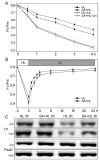
PSII photosensitivity of Arabidopsis thaliana in the presence or absence of SA pretreatment during high light illumination. (A) Untreated (−) and lincomycin-treated (+) detached leaves were exposed to high-light (HL) conditions (1000 μmol photons m−2·s−1) for 4 h. (B) The photoinhibited detached leaves were recovered at low light (LL) intensity (10 μmol photons m−2·s−1) up to 24 h with regular measurement of Fv/Fm. Values are means ± SD from three independent biological replicates (n = 3). (C) Immunoblot analysis of thylakoid proteins obtained from Arabidopsis thaliana in the presence or absence of SA pretreatment with D1 and PsaD antibodies before (−) and after (+) photoinhibition using a light intensity of 1000 μmol photons m−2·s−1 for 3 h. PsaD was as a loading control.
Figure 5
Figure 5
Immunoblot analyses of thylakoid proteins obtained from Arabidopsis thaliana under high light in the presence or absence of SA pretreatment. (A) Immunoblotting were done using specific antibodies against representative PSI and PSII proteins. (B) Quantitative data for D1, D2, Lhcb5, and PsbS proteins in Arabidopsis thaliana under high light with or without SA pretreatment. Results are presented relative to the amount of the respective control (HL 0h, 100%). Significantly different values are marked with an asterisk (*) at p < 0.05 level (n = 4). HL, high light. SA + HL, high light after SA pretreatment for 3 d. 0–3 h, high light for 0 h, 1 h, and 3 h in the presence or absence of SA pretreatment, respectively.
Figure 6
Figure 6
Thylakoid protein phosphorylation of Arabidopsis thaliana under high light in the presence or absence of SA. (A) Immunoblotting of thylakoid membrane proteins analysis was performed using antiphosphothreonine antibodies. (B) Coomassie blue staining (CBS) of SDS-PAGE was presented in the bottom panel. HL, high light. SA + HL, high light after SA pretreatment for 3 d. 0–3 h, high light for 0 h, 1 h, and 3 h in the presence or absence of SA pretreatment, respectively.
Figure 7
Figure 7
Oxygen evolution and thylakoid membrane complexes analysis of thylakoid proteins obtained from Arabidopsis thaliana under high light in the presence or absence of SA. (A) Oxygen evolution rates of thylakoid membranes were measured at 20 °C with 0.5 mM phenyl-p-benzoquinone under saturating light intensities. The data represent means ± SD (standard deviations) from four independent biological replicates (n = 4). Different lower-case letters indicate significant differences (p < 0.05) according to Duncan’s multiplication range test. (B) BN-PAGE of thylakoid membranes was performed using 5–12.5% acrylamide after solubilization using 1% (w/v) DM. NDH, NAD(P)H dehydrogenase; PS, photosystem; LHC, light-harvesting complex; Cyt b6/f, cytochrome b6/f; mc, megacomplex; sc, super complex. (C) Quantitative data for PSII-LHCII super complexes, LHCII trimer, LHCII assembly, and LHCII monomer in Arabidopsis thaliana under high light with or without SA pretreatment. Results are shown relative to the amount of the respective control (HL 0h, 100%). Significantly different values are marked with an asterisk (*) at p < 0.05 level (n = 4). HL, high light. SA + HL, high light after SA pretreatment for 3 d. 0–3 h, high light for 0 h, 1 h, and 3 h in the presence or absence of SA pretreatment, respectively.
Figure 8
Figure 8
Transmission electron microscope analysis of chloroplasts in Arabidopsis thaliana under high light in the presence or absence of SA. HL 0h, high light for 0 h without SA pretreatment. SA+HL 0h, high light for 0 h with SA pretreatment. HL 3 h, high light for 3 h without SA pretreatment. SA + HL 3 h, high light for 3 h with SA pretreatment. Scale bar represents 1 μm in each figure.

References

    1. Pagliano C., Saracco G., Barber J. Structural, functional and auxiliary proteins of photosystem II. Photosynth. Res. 2013;116:167–188. doi: 10.1007/s11120-013-9803-8. - DOI - PubMed
    1. Adir N., Zer H., Shochat S., Ohad I. Photoinhibition—A historical perspective. Photosynth. Res. 2003;76:343–370. doi: 10.1023/A:1024969518145. - DOI - PubMed
    1. Murata N., Takahashi S., Nishiyama Y., Allakhverdiev S.I. Photoinhibition of photosystem II under environmental stress. Biochim. Biophys. Acta Bioenerg. 2007;1767:414–421. doi: 10.1016/j.bbabio.2006.11.019. - DOI - PubMed
    1. Niyogi K.K., Truong T.B. Evolution of flexible non-photochemical quenching mechanisms that regulate light harvesting in oxygenic photosynthesis. Curr. Opin. Plant Biol. 2013;16:307–314. doi: 10.1016/j.pbi.2013.03.011. - DOI - PubMed
    1. Dall’Osto L., Cazzaniga S., Bressan M., Paleček D., Židek K., Niyogi K.K., Fleming G.R., Zigmantas D., Bassi R. Two mechanisms for dissipation of excess light in monomeric and trimeric light-harvesting complexes. Nat. Plants. 2017;3:1–9. doi: 10.1038/nplants.2017.33. - DOI - PubMed

MeSH terms

LinkOut - more resources