. 2020 Feb 14;20(1):52.
doi: 10.1186/s12886-020-01337-9.

Optical coherence tomography features of retinal lesions in Chinese patients with endogenous Candida endophthalmitis

Affiliations

Optical coherence tomography features of retinal lesions in Chinese patients with endogenous Candida endophthalmitis

Hong Zhuang et al. BMC Ophthalmol. .

Abstract

Background: To evaluate the optical coherence tomography (OCT) features of retinal lesions in Chinese patients with endogenous Candida endophthalmitis (ECE).

Methods: We performed a retrospective review of patients diagnosed with ECE at one medical center. The medical records of the patients including predisposing risk factors, treatment and visual acuity were reviewed. And we focused on the analysis of OCT images of retinal lesions before and after treatment.

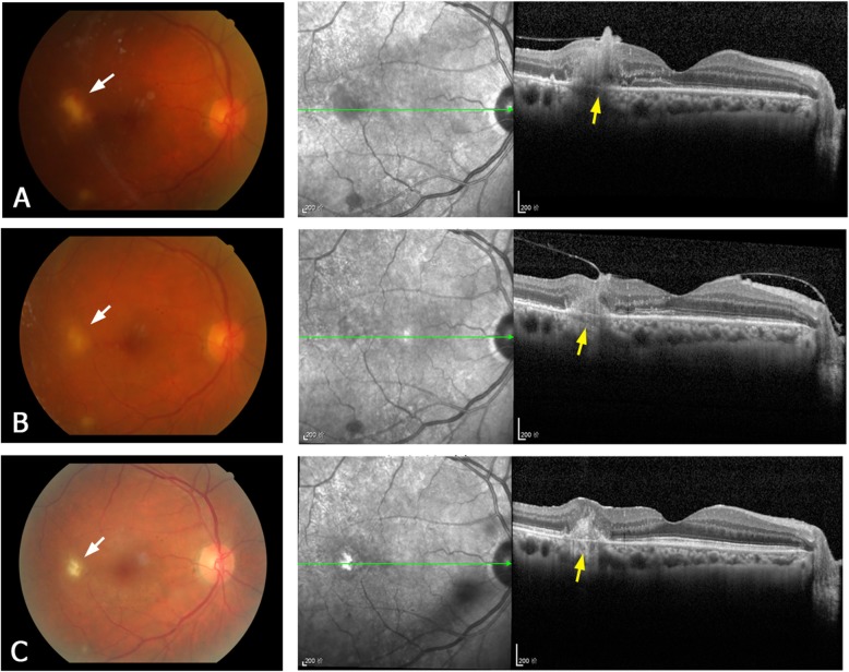
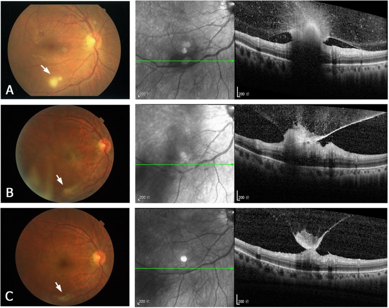
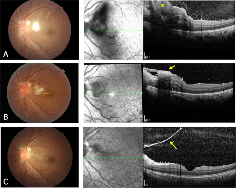
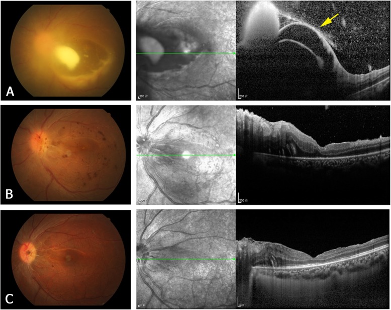
Results: A total of 16 Chinese patients (22 eyes) were included in this study. The most frequent predisposing risk factors were intravenous use of corticosteroids or antibiotics, lithotripsy for urinary calculi, and diabetes. After treatment, visual acuity was improved in 13 (59.1%) of the 22 eyes, and remained the same in the other 9 (40.9%) eyes. Pre-treatment OCT images obtained at presentation were available for 17 of the 22 eyes. Four types of the OCT manifestations of retinal lesions were identified: type 1 (subretinal macular lesions), type 2 (lesions are located in the inner retinal layer), type 3 (lesions involve the full-thickness retina and accompanied with macular edema), type 4 (sub-inner limiting membrane lesions). Pre-treatment OCT imaging of the 17 eyes revealed five as type 1, four as type 2, six as type 3, and two as type 4. After treatment, OCT images revealed epiretinal membrane and subretinal fibrosis as the most common post-treatment complications of ECE. Epiretinal membrane was detected in 2/4 type 2 lesions, in 4/6 type 3 lesions, and in 1/2 type 4 lesions, while subretinal fibrosis was mainly seen in type 1 lesions (4/5). Among the types, visual prognosis was best in eyes with type 2 lesions.

Conclusions: In this case series, the OCT manifestations of retinal lesions in ECE could be classified into four types. The post-treatment OCT manifestations were different in four types of lesions. We preliminarily found that the OCT morphology of retinal lesions was associated with the visual prognosis of ECE.

Keywords: Candida albicans; Endogenous endophthalmitis; Optical coherence tomography; Retinal lesion.


Conflict of interest statement

The authors declare that they have no competing interests.

Figures

Fig. 1
Fig. 1
A representative type 1 lesion of ECE. Fundus photographs (left column) and OCT images (right column) of the right eye of patient 5. a Before treatment. The fundus photograph shows a yellow–white lesion with a small hemorrhage (white arrow) on the temporal side of the fovea. The OCT image shows a macular subretinal lesion originating at the retinal pigment epithelium/choroid layer (yellow arrow) and penetrating into the neurosensory retina. b Two weeks after treatment (intravitreal injection). Shrinkage of the subretinal lesion is apparent on the OCT image. c Five months after treatment. The OCT image shows hyperreflective fibrosis in the subretinal lesion. The inner choroid is also hyperreflective
Fig. 2
Fig. 2
A representative type 2 lesion of ECE. Fundus photographs (left column) and OCT images (right column) of the right eye of patient 10. a Before treatment. The fundus photograph shows a white round lesion (approximately one-half disc diameter) near the infratemporal vascular arcade (white arrow). The OCT image shows a lesion in the inner retinal layer, invading the posterior vitreous. No intra- or subretinal fluid is present. b One month after treatment (intravitreal injection). The OCT image shows shrinkage of the retinal lesion. c Three months after treatment. In the OCT image, regression of the retinal lesion and formation of epiretinal membrane are apparent
Fig. 3
Fig. 3
A representative type 3 lesion of ECE. Fundus photographs (left column) and OCT images (right column) of the left eye of patient 3. a Before treatment. The fundus photograph shows a white fluffy lesion at the posterior pole. The OCT image shows a highly reflective lesion (yellow asterisk) involving the full-thickness retina and protruding into the vitreous. Macular edema is present with subretinal fluid. The hyperreflective dots in the posterior vitreous are infiltrating inflammatory cells. b One week after treatment (vitrectomy). The OCT image shows shrinkage of the retinal lesion, formation of epiretinal membrane (yellow arrow) and reduction of the macular edema. c Three months after treatment. The retinal lesion almost resolves, and OCT shows a residual pre-retinal membrane (yellow arrow) close to the optic disc
Fig. 4
Fig. 4
A representative type 4 lesion of ECE. Fundus photographs (left column) and OCT images (right column) of the left eye of patient 6. a Before treatment. The fundus photograph shows a large lesion at the posterior pole. The OCT image shows detachment of the ILM (yellow arrow) and the sub-ILM lesion breaking through the ILM into the vitreous. b Two weeks after treatment (vitrectomy). In the OCT image, the retinal lesion is no longer apparent, but the nasal retinal edema remains. c Three months after treatment. The OCT image shows reduction of the retinal edema and improvement of the macular foveal structure

References

    1. Okada AA, Johnson RP, Liles WC, D'Amico DJ, Baker AS. Endogenous bacterial endophthalmitis. Report of a ten-year retrospective study. Ophthalmology. 1994;101(5):832–838. doi: 10.1016/S0161-6420(13)31255-X. - DOI - PubMed
    1. Shrader SK, Band JD, Lauter CB, Murphy P. The clinical spectrum of endophthalmitis: incidence, predisposing factors, and features influencing outcome. J Infect Dis. 1990;162(1):115–120. doi: 10.1093/infdis/162.1.115. - DOI - PubMed
    1. Ramakrishnan R, Bharathi MJ, Shivkumar C, Mittal S, Meenakshi R, Khadeer MA, et al. Microbiological profile of culture-proven cases of exogenous and endogenous endophthalmitis: a 10-year retrospective study. Eye (Lond) 2009;23(4):945–956. doi: 10.1038/eye.2008.197. - DOI - PubMed
    1. Wong JS, Chan TK, Lee HM, Chee SP. Endogenous bacterial endophthalmitis: an east Asian experience and a reappraisal of a severe ocular affliction. Ophthalmology. 2000;107(8):1483–1491. doi: 10.1016/S0161-6420(00)00216-5. - DOI - PubMed
    1. Lim HW, Shin JW, Cho HY, Kim HK, Kang SW, Song SJ, et al. Endogenous endophthalmitis in the Korean population: a six-year retrospective study. Retina. 2014;34(3):592–602. doi: 10.1097/IAE.0b013e3182a2e705. - DOI - PubMed

MeSH terms