

Intraoperative 68Ga-PSMA Cerenkov Luminescence Imaging for Surgical Margins in Radical Prostatectomy: A Feasibility Study
- PMID: 32060212
- PMCID: PMC7539648
- DOI: 10.2967/jnumed.119.240424
Intraoperative 68Ga-PSMA Cerenkov Luminescence Imaging for Surgical Margins in Radical Prostatectomy: A Feasibility Study
Abstract
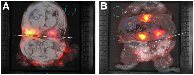
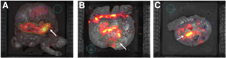
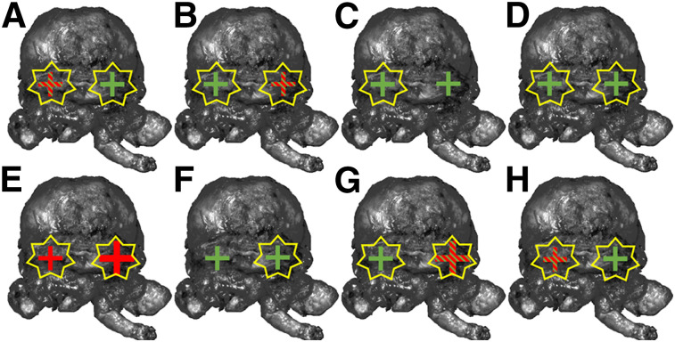
Our objective was to assess the feasibility and accuracy of Cerenkov luminescence imaging (CLI) for assessment of surgical margins intraoperatively during radical prostatectomy. Methods: A single-center feasibility study included 10 patients with high-risk primary prostate cancer (PC). 68Ga-prostate-specific membrane antigen (PSMA) PET/CT scans were performed followed by radical prostatectomy and intraoperative CLI of the excised prostate. In addition to imaging the intact prostate, in the first 2 patients the prostate gland was incised and imaged with CLI to visualize the primary tumor. We compared the tumor margin status on CLI to postoperative histopathology. Measured CLI intensities were determined as tumor-to-background ratio. Results: Tumor cells were successfully detected on the incised prostate CLI images as confirmed by histopathology. Three of 10 men had histopathologically positive surgical margins (PSMs), and 2 of 3 PSMs were accurately detected on CLI. Overall, 25 (72%) of 35 regions of interest proved to visualize a tumor signal according to standard histopathology. The median tumor radiance in these areas was 11,301 photons/s/cm2/sr (range, 3,328-25,428 photons/s/cm2/sr), and median tumor-to-background ratio was 4.2 (range, 2.1-11.6). False-positive signals were seen mainly at the prostate base, with PC cells overlaid by benign tissue. PSMA immunohistochemistry revealed strong PSMA staining of benign gland tissue, which impacts measured activities. Conclusion: This feasibility showed that 68Ga-PSMA CLI is a new intraoperative imaging technique capable of imaging the entire specimen's surface to detect PC tissue at the resection margin. Further optimization of the CLI protocol, or the use of lower-energy imaging tracers such as 18F-PSMA, is required to reduce false-positives. A larger study will be performed to assess diagnostic performance.
Keywords: Cerenkov luminescence imaging; margin assessment; prostate cancer; radical prostatectomy; radioguided surgery.
© 2020 by the Society of Nuclear Medicine and Molecular Imaging.
Figures
Comment in
-
Cerenkov Luminescence Imaging for Surgical Margins in Radical Prostatectomy: A Surgical Perspective.J Nucl Med. 2020 Oct;61(10):1498-1499. doi: 10.2967/jnumed.120.243303. Epub 2020 Jun 26. J Nucl Med. 2020. PMID: 32591492 No abstract available.
References
-
- EAU guidelines on upper urinary tract urothelial carcinoma. Arnhem, The Netherlands: EAU Guidelines Office; 2019. https://uroweb.org/wp-content/uploads/EAU-Guidelines-on-Upper-urinary-Tr.... Accessed September 4, 2020.
-
- Patel VR, Sivaraman A, Coelho RF, et al. Pentafecta: a new concept for reporting outcomes of robot-assisted laparoscopic radical prostatectomy. Eur Urol. 2011;59:702–707. - PubMed
-
- Kozal S, Peyronnet B, Cattarino S, et al. Influence of pathological factors on oncological outcomes after robot-assisted radical prostatectomy for localized prostate cancer: results of a prospective study. Urol Oncol. 2015;33:330.e1–330.e7. - PubMed
-
- Rogers CG, Khan MA, Craig Miller M, Veltri RW, Partin AW. Natural history of disease progression in patients who fail to achieve an undetectable prostate-specific antigen level after undergoing radical prostatectomy. Cancer. 2004;101:2549–2556. - PubMed
MeSH terms
Substances
LinkOut - more resources
Full Text Sources
Medical
Research Materials
Miscellaneous