. 2020 Feb 14;10(1):2696.
doi: 10.1038/s41598-020-59665-1.

Novel Ocellatin Peptides Mitigate LPS-induced ROS Formation and NF-kB Activation in Microglia and Hippocampal Neurons

Affiliations

Novel Ocellatin Peptides Mitigate LPS-induced ROS Formation and NF-kB Activation in Microglia and Hippocampal Neurons

Nayara A Sousa et al. Sci Rep. .

Abstract

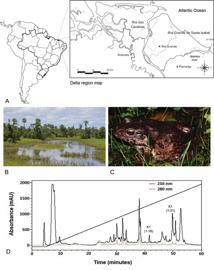
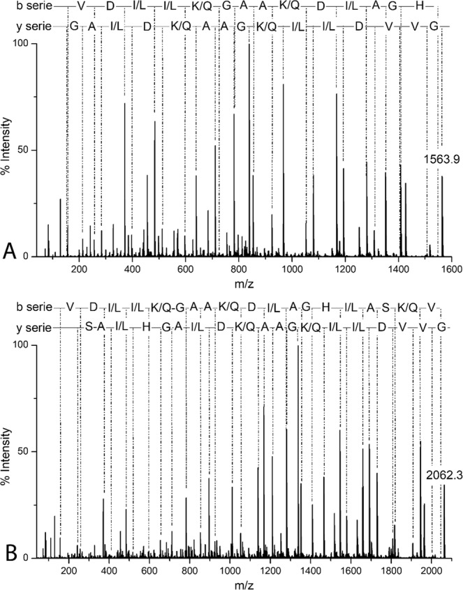
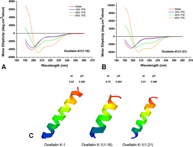
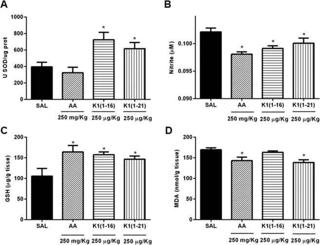
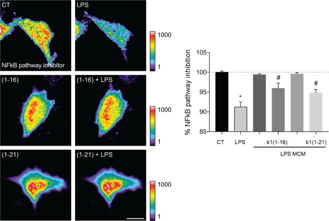
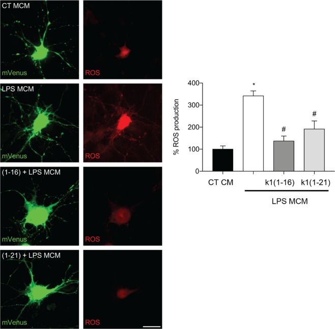
Cutaneous secretions of amphibians have bioactive compounds, such as peptides, with potential for biotechnological applications. Therefore, this study aimed to determine the primary structure and investigate peptides obtained from the cutaneous secretions of the amphibian, Leptodactylus vastus, as a source of bioactive molecules. The peptides obtained possessed the amino acid sequences, GVVDILKGAAKDLAGH and GVVDILKGAAKDLAGHLASKV, with monoisotopic masses of [M + H]± = 1563.8 Da and [M + H]± = 2062.4 Da, respectively. The molecules were characterized as peptides of the class of ocellatins and were named as Ocellatin-K1(1-16) and Ocellatin-K1(1-21). Functional analysis revealed that Ocellatin-K1(1-16) and Ocellatin-K1(1-21) showed weak antibacterial activity. However, treatment of mice with these ocellatins reduced the nitrite and malondialdehyde content. Moreover, superoxide dismutase enzymatic activity and glutathione concentration were increased in the hippocampus of mice. In addition, Ocellatin-K1(1-16) and Ocellatin-K1(1-21) were effective in impairing lipopolysaccharide (LPS)-induced reactive oxygen species (ROS) formation and NF-kB activation in living microglia. We incubated hippocampal neurons with microglial conditioned media treated with LPS and LPS in the presence of Ocellatin-K1(1-16) and Ocellatin-K1(1-21) and observed that both peptides reduced the oxidative stress in hippocampal neurons. Furthermore, these ocellatins demonstrated low cytotoxicity towards erythrocytes. These functional properties suggest possible to neuromodulatory therapeutic applications.


Conflict of interest statement

The authors declare no competing interests.

Figures

Figure 1
Figure 1
The largest delta in open seas in the Americas and the third largest in the world, the Rio Parnaiba Delta covers 70 islands within its 2,700 km2 area, that includes dunes, mangrove forest, and streams (A) (ArcMap v10.3, http://desktop.arcgis.com/en/arcmap/10.3). A temporary pond located in the city of Ilha Grande, State of Piaui. This habitat is home to L. vastus in this region (B). Adult male specimen of L. vastus (Photo: Jose Roberto S. A. Leite) (C). Reverse-phase HPLC chromatogram of the crude extract from L. vastus skin secretion. Fractions containing Ocellatin-K1(1–16) and Ocellatin-K1(1–21) are shown (D).
Figure 2
Figure 2
MS/MS spectra for de novo sequencing of Ocellatin-K1(1–16), [M + H] ±  = 1563.9 Da (A) and Ocellatin-K1(1–21), [M + H]±  = 2062.3 Da (B) acquired using an MALDI-TOF/TOF UltraFlex Xtreme mass spectrometer.
Figure 3
Figure 3
Circular dichroism of peptides in aqueous solution and in 2,2,2-TFE. (A) Ocellatin-K1(1–16) and (B) Ocellatin-K1(1–21). (C) 3D structural model predictions of the ocellatins from Leptodactylus vastus (this study) compared to those of Ocellatin-K1 from Leptodactylus knudseni (Knudsen’s thin-toed frog).
Figure 4
Figure 4
Assessment of anti-microbial activity of Ocellatin-K1(1–16) and Ocellatin-K1(1–21) by MIC assays against E. coli (A) and S. aureus (B) strains in a range of concentrations from 31.25 to 1000 μg/mL. The tests were performed in a single assay in triplicate. The results are expressed as mean ± SEM. *p < 0.05 vs. control group; **p < 0.01 vs. control group; ***p < 0.001 vs. control group; **** p < 0.0001 vs. control group. ANOVA and Sidak test. Abbreviations: K1(1–16): Ocellatin-K1(1–16); K1(1–21): Ocellatin-K1(1–21); OD: optical density.
Figure 5
Figure 5
Oxidative parameters in hippocampus of mice acutely treated with Ocellatin-K1(1–16) and Ocellatin-K1(1–21). (A) SOD relative enzymatic activity, (B) nitrite content, (C) GSH, and (D) MDA concentration. Ascorbic acid was used as a standard antioxidant. The results are expressed as mean ± SEM of a minimum of six animals per group. *p < 0.05 vs. saline group employing ANOVA and Newman–Keuls test. Abbreviations: AA: ascorbic acid; GSH: reduced glutathione; K1(1–16): Ocellatin-K1(1–16); K1(1–21): Ocellatin-K1(1–21); MDA: malondialdehyde; SAL: saline; SOD: superoxide dismutase.
Figure 6
Figure 6
Ocellatin-K1(1–16) and Ocellatin-K1(1–21) prevented the LPS-induced NF-kB activation in living microglia. CHME3 human microglial cells expressing the biosensor of NF-kB pathway inhibitor were incubated with 100 μM Ocellatin-K1(1–16) or Ocellatin-K1(1–21) and challenged with 1 μg/mL LPS. Microglia were incubated only with Ocellatin-K1(1–16) and Ocellatin-K1(1–21) and compared with saline control group. Time-lapse fluorescence intensities for the NF-kB pathway inhibitor biosensor are shown (n = 17–19 cells pooled across two different experiments). The results are expressed as mean ± SEM. #p < 0.01 vs. LPS group; *p < 0.001 vs. CT not treated with LPS employing one-way ANOVA with Bonferroni post-test. Abbreviations: CT: control; LPS: lipopolysaccharide.
Figure 7
Figure 7
Ocellatin-K1(1–16) and Ocellatin-K1(1–21) protect hippocampal neurons from oxidative stress induced by LPS-treated-microglial conditioned media. Representative confocal images and quantification of ROS production in hippocampal neurons incubated with conditioned medium from microglia subjected to LPS-induced and 100 μM Ocellatin-K1(1–16) or Ocellatin-K1(1–21). Images show neurons expressing mVenus (green) and the Hyper Red ROS biosensor (red). The results are expressed as mean ± SEM calculated from 3 different cultures. *p < 0.001 vs. LPS group; #p < 0.001 vs. CT employing one-way ANOVA with the Bonferroni post-test. Abbreviations: CT: control; LPS: lipopolysaccharide; MCM: microglia conditioned medium.
Figure 8
Figure 8
Haemolytic activity of Ocellatin-K1(1–16) and Ocellatin-K1(1–21) in human erythrocytes at concentrations ranging from 7.8 to 500 µg/mL. A positive control was determined using a 10% solution of Triton X-100. ANOVA and t-test were used for statistical analysis.
Figure 9
Figure 9
AFM representative images showing the morphology and roughness of human erythrocytes. (A) Morphology of untreated erythrocytes (control group) and after exposure to the Ocellatin-K1(1–16) (B,D,F at 250, 500, and 1000 µg/mL, respectively) and to the Ocellatin-K1(1–21) (C,E,G at 250, 500, and 1000 µg/mL, respectively). (H) Average roughness of human erythrocytes untreated (control) and treated with Ocellatin-K1(1–16) and Ocellatin-K1(1–21) at concentrations ranging from 250 to 1000 µg/mL. *p < 0.0001 vs. control group using ANOVA and t-test. Abbreviations: K1(1–16): Ocellatin-K1(1–16); K1(1–21): Ocellatin-K1(1–21).

References

    1. Brunetti AE, et al. Cleavage of peptides from amphibian kin revealed by combining analysis of gland secretion and in situ MALDI imaging mass spectrometry. ACS omega. 2018;3:5426–5434. doi: 10.1021/acsomega.7b02029. - DOI - PMC - PubMed
    1. Liu C, et al. Frog skins keep redox homeostasis by antioxidant peptides with rapid radical scavenging ability. Free Radic. Biol. Med. 2010;48:1173–1181. doi: 10.1016/j.freeradbiomed.2010.01.036. - DOI - PubMed
    1. Yang H, et al. Antioxidant peptidomics reveals novel skin antioxidant system. Mol. Cell Proteomics: MCP. 2009;8:571–583. doi: 10.1074/mcp.M800297-MCP200. - DOI - PMC - PubMed
    1. Cao X, et al. Cathelicidin-OA1, a novel antioxidant peptide identified from an amphibian, accelerates skin wound healing. Sci. Rep. 2018;8:943. doi: 10.1038/s41598-018-19486-9. - DOI - PMC - PubMed
    1. Scheele BC, et al. Amphibian fungal panzootic causes catastrophic and ongoing loss of biodiversity. Science. 2019;363:1459–1463. doi: 10.1126/science.aav0379. - DOI - PubMed

Publication types

MeSH terms