

Prevalence of abnormal findings in 230 knees of asymptomatic adults using 3.0 T MRI
- PMID: 32060622
- PMCID: PMC7237395
- DOI: 10.1007/s00256-020-03394-z
Prevalence of abnormal findings in 230 knees of asymptomatic adults using 3.0 T MRI
Abstract
Objective: To identify abnormalities in asymptomatic sedentary individuals using 3.0 Tesla high-resolution MRI.
Materials and methods: The cohort comprised of 230 knees of 115 uninjured sedentary adults (51 males, 64 females; median age: 44 years). All participants had bilateral knee 3.0 T MRIs. Two senior musculoskeletal radiologists graded all intraarticular knee structures using validated scoring systems. Participants completed Knee Injury and Osteoarthritis Outcome Score questionnaires at the time of the MRI scan.
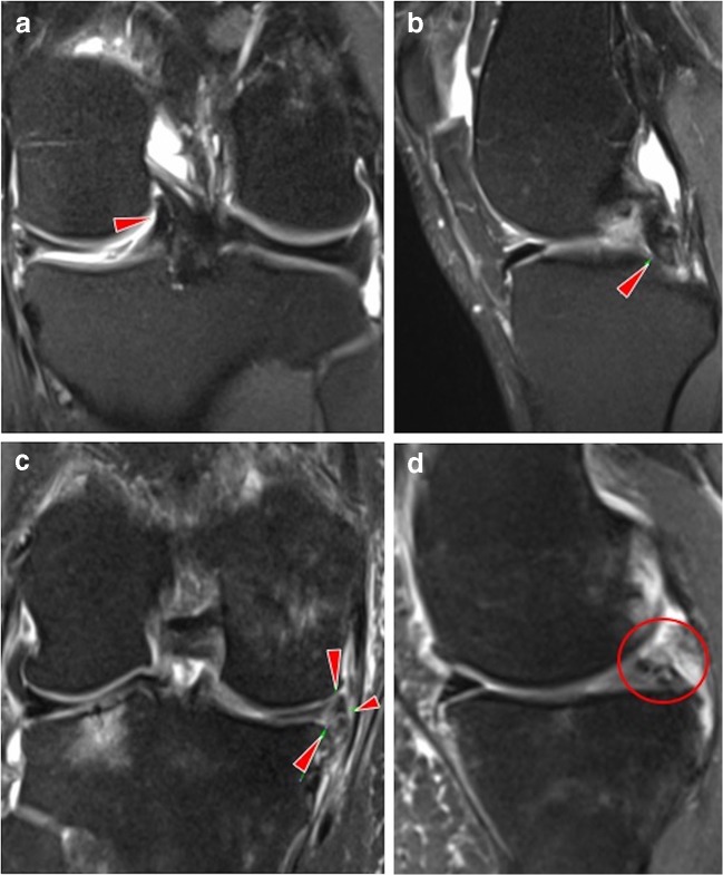
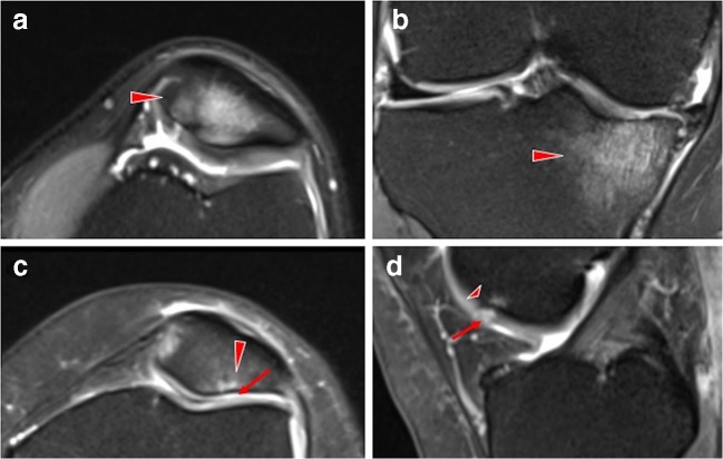
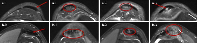
Results: MRI showed abnormalities in the majority (97%) of knees. Thirty percent knees had meniscal tears: horizontal (23%), complex (3%), vertical (2%), radial (2%) and bucket handle (1%). Cartilage and bone marrow abnormalities were prevalent at the patellofemoral joint (57% knees and 48% knees, respectively). Moderate and severe cartilage lesions were common, in 19% and 31% knees, respectively, while moderate and severe bone marrow oedema in 19% and 31% knees, respectively. Moderate-intensity lesion in tendons was found in 21% knees and high-grade tendonitis in 6% knees-the patellar (11% and 2%, respectively) and quadriceps (7% and 2%, respectively) tendons being most affected. Three percent partial ligamentous ruptures were found, especially of the anterior cruciate ligament (2%).
Conclusion: Nearly all knees of asymptomatic adults showed abnormalities in at least one knee structure on MRI. Meniscal tears, cartilage and bone marrow lesions of the patellofemoral joint were the most common pathological findings. Bucket handle and complex meniscal tears were reported for the first time in asymptomatic knees.
Keywords: Elderly; Knee injuries; Pain-free; Radiology.
Conflict of interest statement
The authors declare that they have no competing interests.
Figures
References
-
- Beattie KA, Boullos P, Pui M, O’Neill J, Inglis D, Webber CE, et al. Abnormalities identified in the knees of asymptomatic volunteers using peripheral magnetic resonance imaging. Osteoarthr Cartil. 2005;13(3):181–186. - PubMed
-
- Guymer E, Baranyay F, Wluka AE, Hanna F, Bell RJ, Davis SR, et al. A study of the prevalence and associations of subchondral bone marrow lesions in the knees of healthy, middle-aged women. Osteoarthr Cartil. 2007;15(12):1437–1442. - PubMed
MeSH terms
Grants and funding
LinkOut - more resources
Full Text Sources
Medical
