Review
. 2020 Jun;12(3):719-730.
doi: 10.1007/s12551-020-00636-1. Epub 2020 Feb 15.

Tracking the homeostasis of second messenger cyclic-di-GMP in bacteria

Affiliations
Review

Tracking the homeostasis of second messenger cyclic-di-GMP in bacteria

Anushya Petchiappan et al. Biophys Rev. 2020 Jun.

Abstract

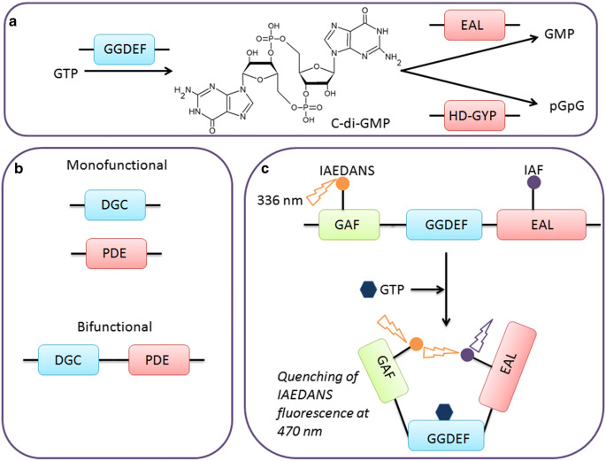
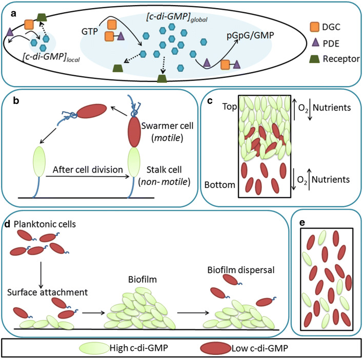
Cyclic-di-GMP (c-di-GMP) is an important second messenger in bacteria which regulates the bacterial transition from motile to sessile phase and also plays a major role in processes such as cell division, exopolysaccharide synthesis, and biofilm formation. Due to its crucial role in dictating the bacterial phenotype, the synthesis and hydrolysis of c-di-GMP is tightly regulated via multiple mechanisms. Perturbing the c-di-GMP homeostasis affects bacterial growth and survival, so it is necessary to understand the underlying mechanisms related to c-di-GMP metabolism. Most techniques used for estimating the c-di-GMP concentration lack single-cell resolution and do not provide information about any heterogeneous distribution of c-di-GMP inside cells. In this review, we briefly discuss how the activity of c-di-GMP metabolising enzymes, particularly bifunctional proteins, is modulated to maintain c-di-GMP homeostasis. We further highlight how fluorescence-based methods aid in understanding the spatiotemporal regulation of c-di-GMP signalling. Finally, we discuss the blind spots in our understanding of second messenger signalling and outline how they can be addressed in the future.

Keywords: Biofilm; Biosensor; C-di-GMP; FRET; Riboswitch; Second messenger.


Conflict of interest statement

The authors declare that they have no conflict of interest.

Figures

Fig. 1
Fig. 1
C-di-GMP metabolism in bacteria. a Synthesis and hydrolysis of c-di-GMP. GGDEF-domain-containing enzymes synthesize c-di-GMP from two molecules of GTP. It is hydrolysed by EAL-domain proteins and HD-GYP domain proteins to GMP and pGpG respectively. b Domain architecture of DGCs and PDEs. Monofunctional enzymes have either synthesis (DGC) or hydrolysis (PDE) activity wherase bifunctional enzymes possess both activities. c Analysis of domain dependence in bifunctional proteins by FRET. M. smegmatis DcpA contains GGDEF and EAL domains along with a regulatoyr EAL domain. The Cys124 in GAF domain and Cys579 in EAL domain are labelled with fluorophore IAEDANS (donor) and IAF (acceptor). Upon binding of its substrate GTP, there is a compaction of protein structure leading to the fluorophores coming proximal to one another. This leads to quenching of the IAEDANS fluorophore which can be observed as reduction in fluorophore intensity. The excitation and emission wavelength used are 336 nm and 470 nm
Fig. 2
Fig. 2
Heterogeneity in c-di-GMP distribution in bacteria. a Spatial sequestration of c-di-GMP modules allows for global and local pools of c-di-GMP which affect bacterial phenotype by binding their receptors. b During the cell division in C. crescentus, the mother cell gives rise to daughter cells with an asymmetric distribution of c-di-GMP. The swarmer cell and the stalk cell contain low and high level of c-di-GMP immediately after division and this dictates their motility. c A cross-section of a biofilm is shown with a spatial distribution of c-di-GMP. Due to the inherent differences in availability of oxygen and nutrients, the expression of PDE varies leading to differences in c-di-GMP concentration. d There is a temporal regulation of c-di-GMP during different stages of biofilm formation. The planktonic cells have low c-di-GMP concentration in contrast to biofilm cells. The c-di-GMP levels increase during surface adhesion and decrease during dispersal. e Isogenic cells in a culture display a bimodal distribution of c-di-GMP
Fig. 3
Fig. 3
Fluorescence-based methods for monitoring c-di-GMP homeostasis. a Reporter-based construct. The expression of a fluorescent reporter protein is dependent on the c-di-GMP-responsive promoter and hence the fluorescence intensity can be monitored to estimate the c-di-GMP concentration. b Riboswitch-based reporter. The expression of a fluorescent reporter protein is dependent on the c-di-GMP-binding riboswitch. The fluorescence is proportional to the c-di-GMP level. c A c-di-GMP-binding riboswitch is fused to a DFHBI-binding aptamer sequence. The binding of c-di-GMP leads to an altered conformation of the riboswitch which can subsequently bind DFHBI leading to fluorescence. d. Split-GFP reporter. FimX and PilZ are expressed as translational fusions with one of three non-functional portions of GFP, GFP11, and GFP10 respectively. GFP1–9 is expressed separately. C-di-GMP increases the interaction between FimX and PilZ. GFP11 and GFP10 can subsequently reconstitute with GFP1–9 to form a fluorescent GFP. The GFP fluorescence is an indicator of c-di-GMP concentration. e FRET-based biosensor. A c-di-GMP-binding protein is sandwiched between YFP and CFP. The binding of c-di-GMP causes structural alteration bringing YFP and CFP in close proximity. Excitation of YFP leads to FRET and the FRET signal can be measured to estimate c-di-GMP concentration. f CSL-BRET-based biosensor. A fluorescent protein (Venus) is expressed fused to a c-di-GMP-binding protein, which is fused between two non-functional parts of luciferase. In the presence of c-di-GMP, there is a structural alteration in the c-di-GMP-binding protein which causes reconstitution of the split luciferase. The luciferase converts its substrate coelenterazine-h to colelentramide-h with concomitant light emission which is captured by Venus for fluorescence

References

    1. Abel S, Bucher T, Nicollier M, Hug I, Kaever V, Zur Wiesch PA, Jenal U (2013) Bi-modal distribution of the second messenger c-di-GMP controls cell fate and asymmetry during the Caulobacter cell cycle PLoS Genet 9:e1003744 - PMC - PubMed
    1. Amikam D, Weinhouse H, Galperin MY (2010) Moshe Benziman and the discovery of cyclic di-GMP the second messenger cyclic Di-GMP
    1. Bähre H, Kaever V (2017) Identification and quantification of cyclic di-guanosine monophosphate and its linear metabolites by reversed-phase LC-MS/MS. In: c-di-GMP signaling. Springer, pp 45-58 - PubMed
    1. Bedrunka P, Graumann PL. New functions and subcellular localization patterns of c-di-GMP components (GGDEF domain proteins) in B. subtilis. Front Microbiol. 2017;8:794–794. doi: 10.3389/fmicb.2017.00794. - DOI - PMC - PubMed
    1. Bharati BK, Sharma IM, Kasetty S, Kumar M, Mukherjee R, Chatterji D (2012) A full-length bifunctional protein involved in c-di-GMP turnover is required for long-term survival under nutrient starvation in Mycobacterium smegmatis Microbiology 158:1415–1427 doi:10.1099/mic.0.053892-0 - PubMed

LinkOut - more resources