Review
. 2020 May;158(7):1948-1966.e1.
doi: 10.1053/j.gastro.2020.01.050. Epub 2020 Feb 13.

Circadian Rhythms in the Pathogenesis and Treatment of Fatty Liver Disease

Affiliations
Review

Circadian Rhythms in the Pathogenesis and Treatment of Fatty Liver Disease

Anand R Saran et al. Gastroenterology. 2020 May.

Abstract

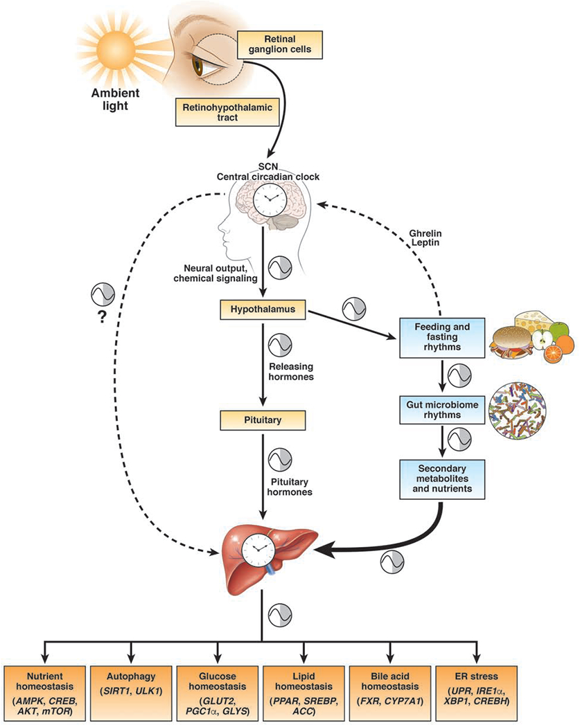
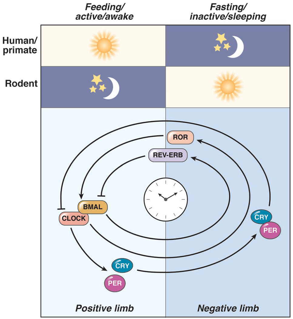
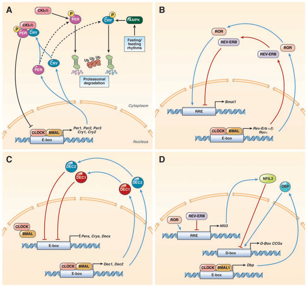
Circadian clock proteins are endogenous timing mechanisms that control the transcription of hundreds of genes. Their integral role in coordinating metabolism has led to their scrutiny in a number of diseases, including nonalcoholic fatty liver disease (NAFLD). Discoordination between central and peripheral circadian rhythms is a core feature of nearly every genetic, dietary, or environmental model of metabolic syndrome and NAFLD. Restricting feeding to a defined daily interval (time-restricted feeding) can synchronize the central and peripheral circadian rhythms, which in turn can prevent or even treat the metabolic syndrome and hepatic steatosis. Importantly, a number of proteins currently under study as drug targets in NAFLD (sterol regulatory element-binding protein [SREBP], acetyl-CoA carboxylase [ACC], peroxisome proliferator-activator receptors [PPARs], and incretins) are modulated by circadian proteins. Thus, the clock can be used to maximize the benefits and minimize the adverse effects of pharmaceutical agents for NAFLD. The circadian clock itself has the potential for use as a target for the treatment of NAFLD.

Keywords: Bile Acids; Dyssynchrony; Gut Microbiome; Lipogenesis; Steatohepatitis.


Figures

Figure 1:
Figure 1:. Central and Peripheral Circadian Rhythms.
Retinal ganglion cells detect ambient light and entrain the suprachiasmatic nucleus (SCN). Their projects travel through the retinohypothalamic tract, though indirect signals are also received from the thalamus. The SCN contains the central circadian clock which can modulate other oscillatory networks including sleep/wake, temperature, and feeding/fasting homeostasis through the hypothalamus. In addition, the SCN regulates the circadian release of hypothalamic releasing hormones (e.g. CRH), and thus, pituitary hormones (e.g. ACTH). Hormones released from pituitary gland are one way that the central clock can regulate the peripheral organs. The SCN also regulates peripheral clock through poorly understood neurohumoral mechanisms. SCN can drive feeding/rhythms through the hypothalamus. Fasting induces the release of ghrelin which promotes feeding while feeding induces the release of leptin which promotes satiety, and thus affecting feeding/fasting rhythms. Diet and feeding/fasting rhythms modulate cyclical fluctuations in the gut microbiome, which then creates cyclical changes in secondary metabolite production, such as SCFAs and bile acids. Secondary metabolites and/or bacterial products are necessary for the maintenance of peripheral circadian rhythms. Feeding/fasting rhythms are powerful drivers of hepatic circadian clock. The rhythmic expression of genes in the liver generates rhythmicity in the transcripts and activity of genes involved in critical metabolic processes such as glucose, lipid, and bile acid metabolism, nutrient homeostasis, autophagy and ER stress. Metabolic syndrome occurs in the setting of central and peripheral circadian rhythm dyssynchrony.
Figure 2:
Figure 2:. Overview of Circadian Clock Machinery.
Humans/primates are diurnal animals with the feeding/active period occurs during the light hours. However, mice (and most rodents) are nocturnal, with the feeding/active period occurring in the dark hours. The cellular molecular clock is a series of transcription-translation feedback loops that include the transcriptional acti vators (CLOCK and BMAL1; the positive limb) and repressors (PER and CRY; the negative limb). The PER:CRY complex repress their own activators CLOCK:BMAL1. CLOCK:BMAL1 also promote REV-ERBs and RORs which add another layer of regulation over the circadian clock and regulate hundreds of additional genes.
Figure 3:
Figure 3:. Circadian Clock Machinery.
(A) In mammals, the core molecular clock is comprised of a series of transcription-translation feedback loops that include the transcriptional activators (CLOCK and BMAL1). The CLOCK:BMAL1 heterodimer regulate genes at a specific DNA binding sequence, the E-box to regulate expression of hundreds of genes, including their own repressors (Per1, Per2, Per3, Cry1, and Cry2). Once translated, PERs and CRYs accumulate in the cytoplasm, where they are regulated by CK1ε/δ and AMPK, respectively. If these PER or CRY are individually phosphorylated, they will undergo ubiquitylation (Ub) and proteasomal degradation. However, if PER and CRY form a heterodimer prior to phosphorylation by CK1ε/δ, the three protein complex is transported to the nucleus, where it directly inhibits the CLOCK:BMAL1 heterodimer. (B) CLOCK:BMAL1 also regulates the expression of Rev-Erbs and Rors. Once translated REV-ERBs and RORs bind to the RRE DNA binding sequence but with opposite effects. RORs promote, while REV-ERBs suppress at this DNA binding sequence. Together these clock proteins control the regulation of hundreds of genes including the expression of Bmal1. (C) A third loop of the circadian clock involves DEC1 and DEC2 that are transcriptionally activated by CLOCK:BMAL1. DEC1 and DEC2 proteins migrate back into the nucleus and inhibit Per1 transactivation by competing for E-Box binding. (D) CLOCK:BMAL1 promote the transcription of Dbp, and REV-ERB/ROR regulate the expression of Nfil3. Once translated, DBP and NFIL3 promote or suppress, respectively, gene expression at the D-Box DNA binding sequence. The D-box DNA binding sequence controls expression of hundreds of clock-controlled genes (CCGs).

References

    1. Gachon F, Nagoshi E, Brown SA, et al. The mammalian circadian timing system: from gene expression to physiology. Chromosoma 2004;113:103–12. - PubMed
    1. Yamazaki S, Numano R, Abe M, et al. Resetting central and peripheral circadian oscillators in transgenic rats. Science 2000;288:682–5. - PubMed
    1. Mure LS, Le HD, Benegiamo G, et al. Diurnal transcriptome atlas of a primate across major neural and peripheral tissues. Science 2018;359. - PMC - PubMed
    1. Damiola F, Le Minh N, Preitner N, et al. Restricted feeding uncouples circadian oscillators in peripheral tissues from the central pacemaker in the suprachiasmatic nucleus. Genes Dev 2000;14:2950–61. - PMC - PubMed
    1. Buijs R, Salgado R, Sabath E, et al. Peripheral circadian oscillators: time and food. Prog Mol Biol Transl Sci 2013;119:83–103. - PubMed

Publication types

MeSH terms

Substances