

White matter injury after neonatal encephalopathy is associated with thalamic metabolite perturbations
- PMID: 32062359
- PMCID: PMC7016374
- DOI: 10.1016/j.ebiom.2020.102663
White matter injury after neonatal encephalopathy is associated with thalamic metabolite perturbations
Abstract
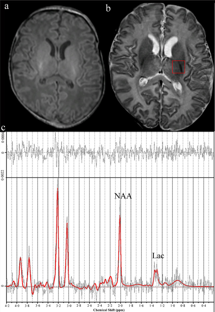
Background: Although thalamic magnetic resonance (MR) spectroscopy (MRS) accurately predicts adverse outcomes after neonatal encephalopathy, its utility in infants without MR visible deep brain nuclei injury is not known. We examined thalamic MRS metabolite perturbations in encephalopathic infants with white matter (WM) injury with or without cortical injury and its associations with adverse outcomes.
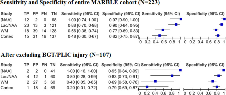
Methods: We performed a subgroup analysis of all infants recruited to the MARBLE study with isolated WM or mixed WM/cortical injury, but no visible injury to the basal ganglia/thalamus (BGT) or posterior limb of the internal capsule (PLIC). We used binary logistic regression to examine the association of MRS biomarkers with three outcomes (i) WM injury score (1 vs. 2/3); (ii) cortical injury scores (0/1 vs. 2/3); and (iii) adverse outcomes (defined as death, moderate/severe disability) at two years (yes/no). We also assessed the accuracy of MRS for predicting adverse outcome.
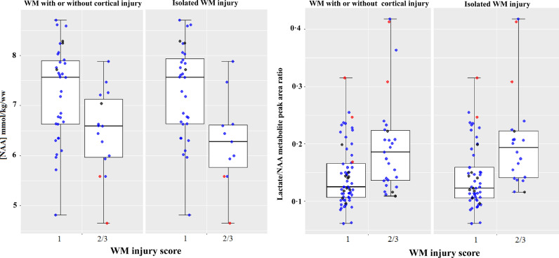
Findings: Of the 107 infants included in the analysis, five had adverse outcome. Reduced thalamic N-acetylaspartate concentration [NAA] (odds ratio 0.4 (95% CI 0.18-0.93)) and elevated thalamic Lactate/NAA peak area ratio (odds ratio 3.37 (95% CI 1.45-7.82)) were significantly associated with higher WM injury scores, but not with cortical injury. Thalamic [NAA] (≤5.6 mmol/kg/wet weight) had the best accuracy for predicting adverse outcomes (sensitivity 1.00 (95% CI 0.16-1.00); specificity 0.95 (95% CI 0.84-0.99)).
Interpretation: Thalamic NAA is reduced in encephalopathic infants without MR visible deep brain nuclei injury and may be a useful predictor of adverse outcomes.
Funding: The National Institute for Health Research (NIHR).
Keywords: Biomarkers; Magnetic resonance imaging; Magnetic resonance spectroscopy; Neonatal encephalopathy; Therapeutic hypothermia.
Copyright © 2020 The Authors. Published by Elsevier B.V. All rights reserved.
Conflict of interest statement
Declaration of Competing Interest We declare no competing interests.
Figures
References
-
- Jacobs S.E., Berg M., Hunt R., Tarnow-Mordi W.O., Inder T.E., Davis P.G. Cooling for newborns with hypoxic ischaemic encephalopathy. Cochrane Database Syst Rev. 2013;(1) - PubMed
-
- Williams C.E., Gunn A.J., Mallard C., Gluckman P.D. Outcome after ischemia in the developing sheep brain: an electroencephalographic and histological study. Ann Neurol. 1992;31(1):14–21. - PubMed
-
- Vannucci R.C. Hypoxic-ischemic encephalopathy. Am J Perinatol. 2000;17(3):113–120. - PubMed
-
- Miller S.P., Ramaswamy V., Michelson D. Patterns of brain injury in term neonatal encephalopathy. J Pediatr. 2005;146(4):453–460. - PubMed
-
- Ancora G., Maranella E., Grandi S. Early predictors of short term neurodevelopmental outcome in asphyxiated cooled infants. a combined brain amplitude integrated electroencephalography and near infrared spectroscopy study. Brain Dev. 2013;35(1):26–31. - PubMed
MeSH terms
Substances
Grants and funding
LinkOut - more resources
Full Text Sources
Medical
