Review
. 2020 May;62(5):563-585.
doi: 10.1007/s00234-019-02357-z. Epub 2020 Feb 15.

The splenium of the corpus callosum: embryology, anatomy, function and imaging with pathophysiological hypothesis

Affiliations
Review

The splenium of the corpus callosum: embryology, anatomy, function and imaging with pathophysiological hypothesis

J Blaauw et al. Neuroradiology. 2020 May.

Abstract

Background and purpose: The splenium of the corpus callosum is the most posterior part of the corpus callosum. Its embryological development, anatomy, vascularization, function, imaging of pathology, possible pathophysiological mechanisms by which pathology may develop and the clinical consequences are discussed.

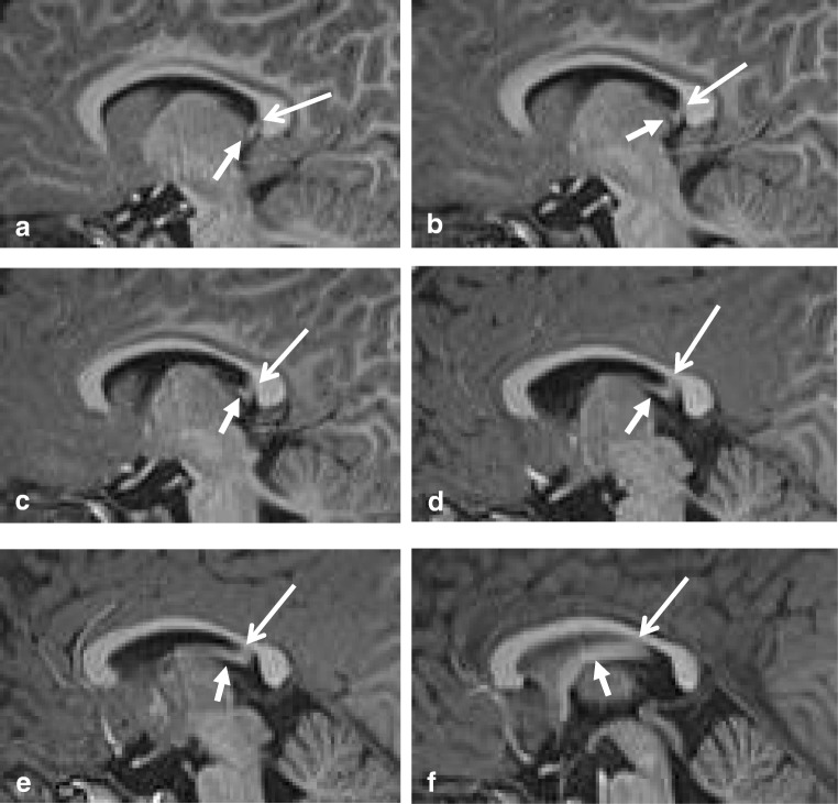
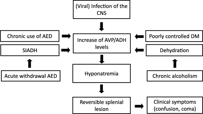
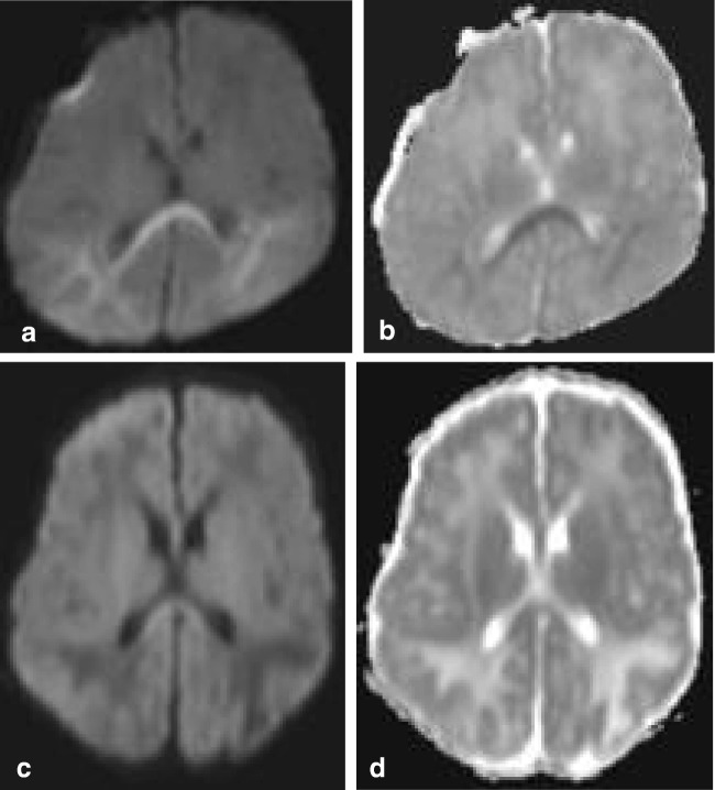
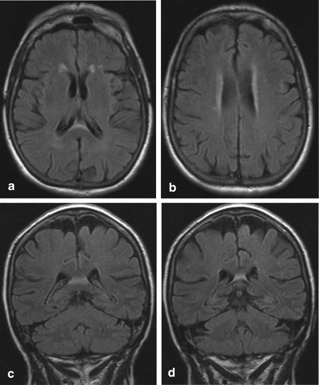
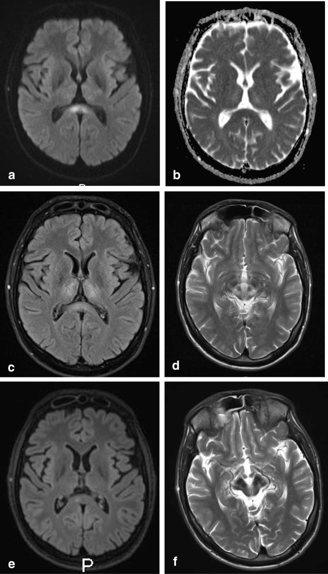
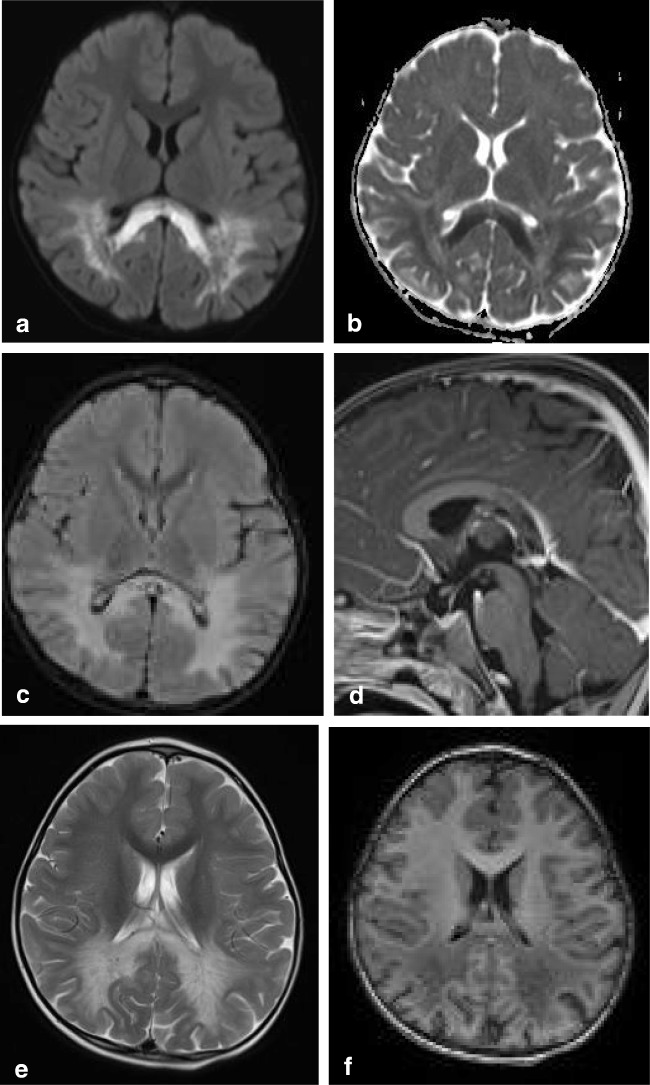
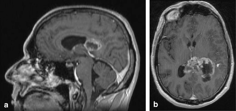
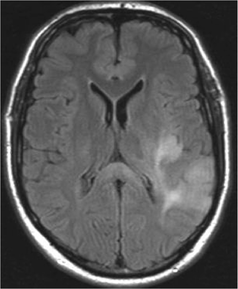
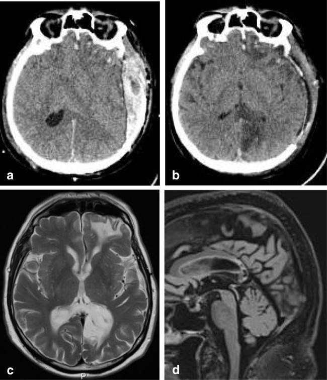
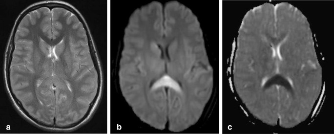
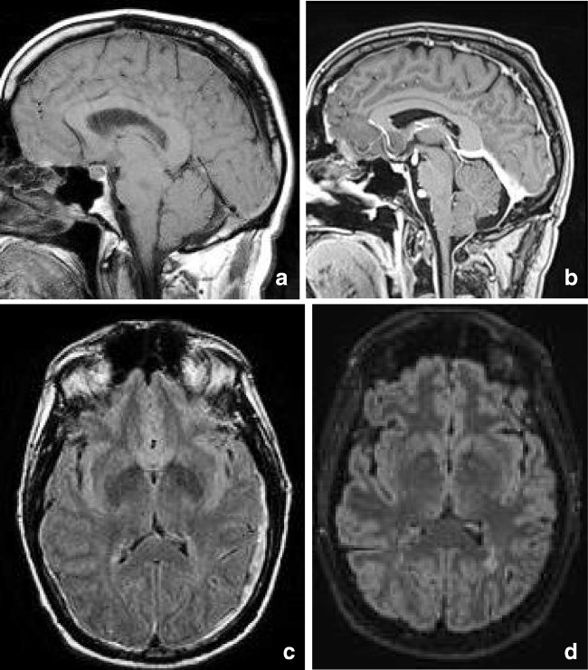
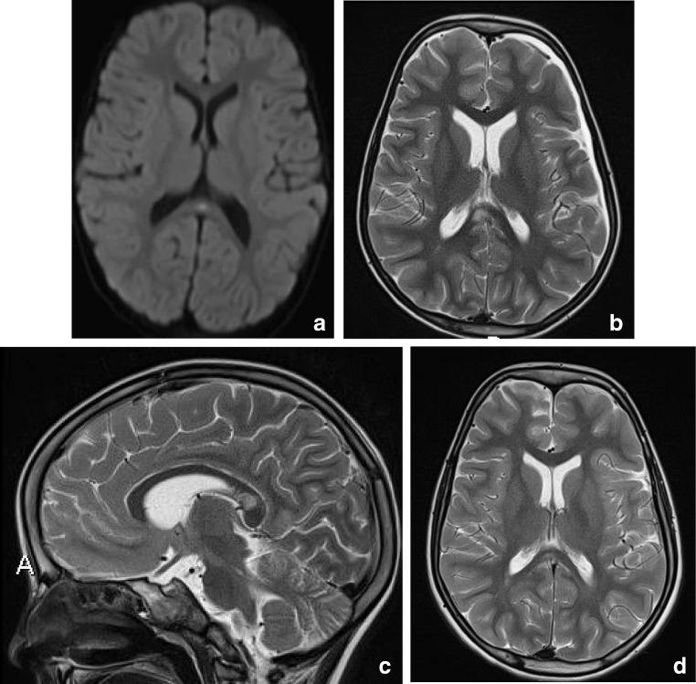
Methods: A literature-based description is provided on development, anatomy and function. MR and CT images are used to demonstrate pathology. The majority of pathology, known to affect the splenium, and the clinical effects are described in three subsections: (A) limited to the splenium, with elaboration on pathophysiology of reversible splenial lesions, (B) pathology in the cerebral white matter extending into or deriving from the splenium, with special emphasis on tumors, and (C) splenial involvement in generalized conditions affecting the entire brain, with a hypothesis for pathophysiological mechanisms for the different diseases.

Results: The development of the splenium is preceded by the formation of the hippocampal commissure. It is bordered by the falx and the tentorium and is perfused by the anterior and posterior circulation. It contains different caliber axonal fibers and the most compact area of callosal glial cells. These findings may explain the affinity of specific forms of pathology for this region. The fibers interconnect the temporal and occipital regions of both hemispheres reciprocally and are important in language, visuospatial information transfer and behavior. Acquired pathology may lead to changes in consciousness.

Conclusion: The development, location, fiber composition and vascularization of the splenium make it vulnerable to specific pathological processes. It appears to play an important role in consciousness.

Keywords: Anatomy; Consciousness; Corpus callosum; MRI; Pathophysiology; Splenium.


Conflict of interest statement

The authors declare that they have no conflict of interest.

Figures

Fig. 1
Fig. 1
Sagittal 3D T1-weighted image (af) with short closed arrow pointing at the fornix and long open arrow indicating the hippocampal commissure, extending from the ventral splenium to the isthmus
Fig. 2
Fig. 2
The flowchart provides a possible mechanism by which a reversible splenial lesion may develop from differents pathological processes leading to hyponatremia
Fig. 4
Fig. 4
Seven-day-old child born at 38 + 5 weeks with a difficult delivery and an APGAR score of 4–5–6 was admitted to another hospital. It was transferred to our hospital because of seizures at 2 days postpartum. On admission, the child had a hypoglycemia (< 0.5 mmol/l) for which glucose infusion was given. DWI (a) and ADC (b) made 5 days after admission showed diffusion restriction in the splenium and in the subcortical white matter of the parietal-occipital lobes. On follow-up DWI (c) and ADC (d) made 16 days after admission, the signal intensity of the splenium normalized, with remaining white matter edema in the parietal lobes and widening of the ventricles, due to global tissueloss
Fig. 5
Fig. 5
Seventy-six-year-old woman. Axial FLAIR (a, b) and coronal FLAIR (c, d). Hyperintense band ventral to the splenium in the region of the hippocampal commissure extending into the fornices
Fig. 6
Fig. 6
Thirty-year-old man. Axial DWI (a) showing a central area of high signal in the splenium, without low signal on ADC (b) and comparable high signal on FLAIR (c), compatible with T2 shine through effect and white matter edema. Axial T2 (d) shows a typical giant panda sign at the level of the mesencephalon-pons. On follow-up 2 years later, the DWI (e) shows a small residual splenial lesion on the right, and axial T2 (f) at the level of the mesencephalon shows atrophy and increase in low signal in the substantia nigra and nucleus ruber
Fig. 7
Fig. 7
Seven-year-old boy with X-linked adrenoleukodystrophy. Axial FLAIR shows hyperintense area located in the ventral splenium
Fig. 8
Fig. 8
Three-year-old boy with Krabbe’s disease. Axial DWI (a) and ADC (b) show extensive abnormal signal with partial diffusion restriction involving the splenium and parietal white matter. Axial FLAIR (c) and sagittal T1 after gadolinium (d) show widened (hypointense) perivascular spaces in the splenium. Axial T2 (e) and inversion recovery (f) show spared axons in the parietal white matter
Fig. 9
Fig. 9
Fifty-year-old male with a glioblastoma multiforme. Sagittal contrast-enhanced T1 (a) and axial T2 (b) show irregular tumor mass with central necrosis and irregular enhancing rim in the splenium, extending into the left parietal white matter
Fig. 10
Fig. 10
Forty-nine-year-old man with a diffuse glioma grade 2. Axial FLAIR shows a diffuse tumor with extension into the splenium, consistent with appearance of gliomatosis cerebri
Fig. 11
Fig. 11
Forty-seven-year-old man with biopsy proven B cell lymphoma. Axial FLAIR (a) shows a space occupying lesion with a slightly hyperintense center and a hypointense rim in the splenium, which enhances vividly after contrast (axial T1 with gadolinium (b)
Fig. 12
Fig. 12
Sixty-four-year-old man with biopsy proven demyelination with some T cell infiltrate and slight secondary axonal injury, consistent with ADEM. Axial DWI (a) and ADC (b) show large splenial lesion with diffusion restriction, without enhancement (not shown). Axial T2 (c) shows lesion in the right cerebellar hemisphere, with central punctiform enhancement (not shown). Swelling of the entire spinal cord shown on sagittal T2 (f, g)
Fig. 13
Fig. 13
Normal control for comparison, 5-year-old boy with headaches. Normally developed CC with comparable thickness of splenium and genu (a). A 3.5-year old boy with a TUBB2A mutation with developmental and motor delay with splenial dysgenesis. Also presenting with developmental disturbance of the pons and cerebellum (b). Five-year-old boy with developmental delay, dysmorphic features, epilepsy, and spastic paraplegia, associated with an AP4S1 gene mutation, with the most severely affected volume restriction of the splenium (c). Five-year-old boy with progressively enlarging skull circumference associated with an mTOR gene mutation. The splenium is dysgenetic, with smaller caliber compared with the genu (d)
Fig. 14
Fig. 14
Two-year-old child. Sagittal T1 shows dysgenesis of the corpus callosum mostly affecting the splenium, with a curvilinear lipoma extending from below the splenium continuing anteriorly, covering the superior surface of the corpus up to the genu (a). Seventy-two-year-old man. Sagittal CT reconstruction shows a sharply demarcated hypodense structure curving posteriorly around a normal appearing splenium, consistent with a lipoma (black arrows). The bright spot inferior to the lipoma is a calcified pineal gland (b)
Fig. 15
Fig. 15
Forty-eight-year-old woman. Axial DWI (a) and ADC (b) show a small area of diffusion restriction in the right splenium (black arrow) with a larger area of edema on the T2 (c). In the corona radiata and frontal white matter, multiple lacunar infarcts are seen (d). Follow-up axial T2 (e) at the level of figure c shows residual focal loss of tissue in right splenium and a similar lesion in left thalamus (black arrow). 3D TOF MRA shows normal cerebral arteries (f)
Fig. 17
Fig. 17
Sixty-six-year-old man. Axial CT with leftsided subdural hematoma and diffuse brain swelling without splenial hypodensity on day of trauma (a); 3 days later, infarction is seen in the left side of the splenium and in the left occipital lobe (b). At follow-up, 7 years later, the patient has no residual cognitive disturbance. Axial T2 (c) and sagittal 3D FLAIR (d) show extreme tissue loss in the same regions
Fig. 16
Fig. 16
Fifteen-year-old girl post high energy  trauma. CT on day of trauma (not shown) showed no abnormalities. Axial T2 MRI (a) at day 6, post trauma, shows involvement of the entire splenium, with restricted diffusion on DWI (b) and ADC map (c). At 6-week follow-up, the patient had severe residual cognitive disturbance
Fig. 18
Fig. 18
Fifty-three-year-old man with intracranial hypotension syndrome. At first admission, sagittal T1 (a) shows an enlarged and downward herniating splenium, with normal signal intensity on the axial FLAIR (c). Also sagging midbrain and bilateral subdural hematoma. Seven years later scanned for left frontal headache, disappearing with reclining. Sagittal T1 after gadolinium (b) shows normalized position brainstem, with decreased enlargement of the splenium. On axial FLAIR (d), no new SDH is seen
Fig. 3
Fig. 3
(a) Three-year-old girl with gastroenteritis by enterovirus. Axial DWI (a) on day 4 shows a small lesion with unsharp border consistent with a RESLES lesion. (b-d) Six-year-old girl with a pneumococcal meningitis following an ear infection. Axial and sagittal T2 at admission (b, c) shows an area of edema in the splenial and dorsal isthmus (no DWI was made). At 5-year follow-up, a small residual lesion is seen on the axial T2 (d)

References

    1. Raybaud C. The corpus callosum, the other great forebrain commissures, and the septum pellucidum: anatomy, development, and malformation. Neuroradiology. 2010;52:447–477. - PubMed
    1. Takanashi J, Tada H, Kuroki H, Barkovich AJ. Delirious behavior in influenza is associated with a reversible splenial lesion. Brain Dev. 2009;31:423–426. - PubMed
    1. Barkovich AJ, Norman D. Anomalies of the corpus callosum: correlation with further anomalies of the brain. Am J Roentgenol. 1988;151:171–179. - PubMed
    1. Rakic P, Yakovlev PI. Development of the corpus callosum and cavum septi in man. J Comp Neurol. 1968;132:45–72. - PubMed
    1. Kier EL, Fulbright RK, Bronen RA. Limbic lobe embryology and anatomy: dissection and MR of the medial surface of the fetal cerebral hemisphere. AJNR Am J Neuroradiol. 1995;16:1847–1853. - PMC - PubMed

MeSH terms