Review
. 2021 May;72(2):258-270.
doi: 10.1177/0846537119895751. Epub 2020 Feb 6.

Mimickers of Hill-Sachs Lesions

Affiliations
Review

Mimickers of Hill-Sachs Lesions

Allison Herring et al. Can Assoc Radiol J. 2021 May.

Abstract

The purpose of this article is to describe the imaging appearance, etiology, clinical features, and treatment of rare presentations of common bone and joint diseases known to mimic Hill-Sachs lesions. Knowledge of uncommonly encountered manifestations of ankylosing spondylitis, rheumatoid arthritis, septic joint, hyperparathyroidism, hydroxyapatite deposition disease, malignant bone tumors, and benign bone cysts which mimic traumatic Hill-Sachs lesions is important for radiologists to guide the clinical care of patients who present with shoulder symptoms.

Keywords: Hill-Sachs lesion; hatchet sign; humerus; imaging; mimicker; shoulder.


Figures
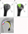
Figure 1.
Figure 1.
Illustrations of proximal humerus anatomy. (A) Anterior and (B) posterior illustrations of the proximal humerus: 1 = greater tuberosity, 2 = lesser tuberosity, 3 = articular surface of the humeral head, 4 = anatomic neck (dotted line), green = typical location of Hill-Sachs lesions and its mimickers. (C) Location of osseous regions at the proximal humerus in relation to the glenohumeral joint in cross-section (coronal plane): red = cortex of the greater tuberosity, blue = bare area, yellow = subchondral bone of the humeral head, purple = rotator cuff tendon, asterisk = intracapsular space of the glenohumeral joint.
Figure 2.
Figure 2.
Hill-Sachs lesions. 53-year-old man after acute fall > 30 feet into water. (A) Anteroposterior (AP) internal rotation shoulder radiograph shows an acute anterior shoulder dislocation. (B) Follow-up AP internal rotation shoulder radiograph shows a Hill-Sachs lesion (arrow) following reduction of the dislocation. 25-year-old man with chronic shoulder instability and history of multiple dislocations. (C) Grashey shoulder radiograph shows a Hill-Sachs lesion (arrow).
Figure 3.
Figure 3.
Ankylosing spondylitis. A 31-year-old man with ankylosing spondylitis complains of multifocal joint pain. (A) Anteroposterior (AP) pelvic radiograph demonstrates complete ankylosis of the right sacroiliac joint (black arrow) and incomplete bony ankylosis of the left sacroiliac joint (white arrow). (B) Lateral cervical spine radiograph demonstrates thin bridging syndesmophytes anteriorly (arrows). (C) AP internal and (D) AP external rotation shoulder radiographs demonstrates a large erosion (arrow) at the posterolateral aspect of the humeral head and greater tuberosity.
Figure 4.
Figure 4.
Rheumatoid Arthritis. A 37-year-old woman with rheumatoid arthritis and chronic shoulder pain. (A) Anteroposterior external rotation shoulder radiograph shows a large erosion (arrow) at the humeral head and greater tuberosity. (B) Oblique coronal short tau inversion recovery-weighted shoulder MR image shows an erosion with associated osteitis / bone marrow edema at the humeral head (long-thin arrow). Rice-bodies (short-thin arrow) and fluid distend the subacromial subdeltoid bursa, and a high-grade articular surface partial tear of the supraspinatus tendon (short-wide arrow) is present.
Figure 5.
Figure 5.
Typical bacterial infection. A 38-year-old man with nonspecific shoulder pain for several months developing septic joint of the shoulder caused by methicillin-sensitive staphylococcus aureus. (A) Anteroposterior neutral shoulder radiograph shows no abnormality on initial presentation. (B) Post-operative Grashey shoulder radiograph following incision and drainage (I&D) surgery 4 months later demonstrates an interval development of diffuse peri-articular osteopenia and a large erosion (arrow) at the junction of the greater tuberosity and humeral head. (C) Oblique coronal short tau inversion recovery-weighted shoulder MR image 2 days prior to I&D surgery shows an erosion with associated bone marrow edema at the lateral aspect of the humeral head (arrow). Diffuse peri-articular soft tissue edema is also present.
Figure 6.
Figure 6.
Typical and atypical bacterial infection. A 50-year-old woman with endocarditis and septic shoulder joint caused by methicillin-resistant staphylococcus aureus. (A) Anteroposterior neutral shoulder radiograph shows an ill-defined erosion at the humeral head and greater tuberosity (arrow). (B) Contrast-enhanced axial CT image shows a large humeral head erosion (arrow) and a large glenohumeral joint effusion (asterisk). A 55-year-old man with disseminated mycobacterium avium-intracellulare complex and septic shoulder joint. (C) Oblique coronal proton density-weighted and (D) oblique sagittal T1-weighted shoulder MR images show a large erosion involving the greater tuberosity and humeral head (arrows) with associated soft tissue abscess (asterisk).
Figure 7.
Figure 7.
Hyperparathyroidism. A 65-year-old woman with long standing end-stage renal disease, secondary hyperparathyroidism and chronic shoulder pain. (A) Anteroposterior internal rotation radiograph and (B) a corresponding unenhanced coronal CT image show a large erosion (long arrow) at the posterolateral aspect of the humeral head, with an associated rotator cuff tear implied by severe narrowing of the acromiohumeral interval. Distal claviclar osteolysis is also present (short arrow).
Figure 8.
Figure 8.
Hydroxyapatite deposition disease. 58-year-old woman with acute on chronic shoulder pain. (A) Oblique coronal short tau inversion recovery-weighted and (B) oblique coronal proton density-weighted shoulder MR images show low signal deposits of calcium hydroxyapatite (long arrow) eroding into the humeral head and greater tuberosity with associated bone marrow edema (asterisk) and adjacent soft tissue edema. Additional foci of calcium hydroxyapatite are associated with the subacromial subdeltoid bursa and rotator cuff (short arrow). (C) A corresponding Grashey shoulder radiograph depicts a focus of calcium hydroxyapatite (arrow).
Figure 9.
Figure 9.
Metastatic disease. A 57-year-old woman with shoulder pain and metastatic cervical carcinoma. (A) Anteroposterior external rotation shoulder radiograph shows a region of ill-defined lytic bone destruction (arrow) involving the posterolateral proximal humerus with a subtle linear lucent step-off at the junction of the humeral head and greater tuberosity suggesting non-displaced pathologic fracture. (B) 18F-FDG PET/CT fused image shows intense FDG avidity in the metastasis at the proximal humerus with a standard uptake value of 17.
Figure 10.
Figure 10.
Benign bone cysts. A 55-year-old man with chronic shoulder pain and decreased range of motion. (A) Grashey shoulder radiograph and (B) a corresponding oblique sagittal T1-weighted shoulder MR image demonstrate a focal bone cyst (arrow) involving the junction of the bare area and greater tuberosity. 57-year-old woman with nonspecific shoulder pain. (C) Grashey shoulder radiograph shows bone cyst formation spanning the junction of the greater tuberosity and humeral head.

References

    1. Jin W, Ryu KN, Park YK, Lee WK, Ko SH, Yang DM. Cystic lesions in the posterosuperior portion of the humeral head on MR arthrography: correlations with gross and histologic findings in cadavers. AJR Am J Roentgenol 2005; 184:1211–1215 - PubMed
    1. Jacobson JA, Girish G, Jiang Y, Resnick D. Radiographic evaluation of arthritis: inflammatory conditions. Radiology 2008; 248:378–389 - PubMed
    1. Kadi R, Milants A, Shahabpour M. Shoulder Anatomy and Normal Variants. J Belg Soc Radiol 2017; 101:3 - PMC - PubMed
    1. Sandstrom CK, Kennedy SA, Gross JA. Acute shoulder trauma: what the surgeon wants to know. Radiographics 2015; 35:475–492 - PubMed
    1. Fox JA, Sanchez A, Zajac TJ, Provencher MT. Understanding the Hill-Sachs Lesion in Its Role in Patients with Recurrent Anterior Shoulder Instability. Curr Rev Musculoskelet Med 2017; 10:469–479 - PMC - PubMed

MeSH terms