Review
. 2020 Jan 29:10:935.
doi: 10.3389/fendo.2019.00935. eCollection 2019.

Regulation of Inflammation Pathways and Inflammasome by Sex Steroid Hormones in Endometriosis

Affiliations
Review

Regulation of Inflammation Pathways and Inflammasome by Sex Steroid Hormones in Endometriosis

Elizabeth García-Gómez et al. Front Endocrinol (Lausanne). .

Abstract

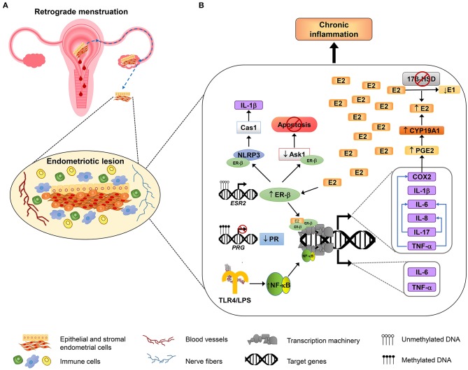
Endometriosis is a gynecological disorder characterized by the growth of endometrial tissue (glands and stroma) outside the uterus, mainly in the peritoneal cavity, ovaries, and intestines. This condition shows estrogen dependency and progesterone resistance, and it has been associated with chronic inflammation, severe pain, and infertility, which negatively affect the quality of life in reproductive women. The molecular mechanisms involved in the pathogenesis of endometriosis are not completely understood; however, inflammation plays a key role in the pathophysiology of the disease, mainly by altering the function of immune cells (macrophages, natural killer, and T cells) and increasing levels of pro-inflammatory mediators in the peritoneal cavity, endometrium, and blood. These immune alterations inhibit apoptotic pathways and promote adhesion and proliferation of endometriotic cells, as well as angiogenesis and neurogenesis in endometriotic lesions. It has been demonstrated that hormonal alterations in endometriosis are related to the inflammatory unbalance in this disease. Particularly, steroid hormones (mainly estradiol) promote the expression and release of pro-inflammatory factors. Excessive inflammation in endometriosis contributes to changes of hormonal regulation by modulating sex steroid receptors expression and increasing aromatase activity. In addition, dysregulation of the inflammasome pathway, mediated by an alteration of cellular responses to steroid hormones, participates in disease progression through preventing cell death, promoting adhesion, invasion, and cell proliferation. Furthermore, inflammation is involved in endometriosis-associated infertility, which alters endometrium receptivity by impairing biochemical responses and decidualization. The purpose of this review is to present current research about the role of inflammasome in the pathogenesis of endometriosis as well as the molecular role of sex hormones in the inflammatory responses in endometriosis.

Keywords: bacteria; endometriosis; estrogen receptor; inflammasome; inflammation; pro-inflammatory factors; progesterone receptor; sex steroid hormones.


Figures

Figure 1
Figure 1
Inflammatory mechanisms involved in endometriosis. An overview about the molecular mechanisms involved in the adhesion, survival, proliferation, inflammation, and neuroangiogenesis of endometriotic lesions. (A) According to the most accepted hypothesis of endometriosis origin, stromal and epithelial cells from shed endometrial tissue reach ovaries or peritoneal cavity by retrograde efflux during menstruation. The developing endometriotic lesion stimulates angiogenesis and nerve development and secretes chemoattractant molecules recruiting high numbers of macrophages (showed in blue) and natural killer cells (indicated in green) with reduced phagocytic and cytolytic activity, respectively. (B) Alterations in DNA methylation lead to the growth and maintenance of endometriotic lesions by increasing the expression of estrogen receptor β (ER-β) and reducing progesterone receptor (PR) expression. Overexpressed ER-β induces the expression of genes that encode several pro-inflammatory molecules, such as IL-1β, IL-6, IL-8, IL-17, TNF-α, and COX2; some of them, in turn, can stimulate the production of other immune molecules (depicted as blue arrows). COX2 promotes an increase of the synthesis of prostaglandin E2 (PGE2), and PGE2 induces aromatase activity (CYP19A1), leading to E2 increased levels. E2 elevated levels are sustained by reduced expression of 17β-HSD, an enzyme that catalyzes the conversion of E2 to estrone (E1), and that is induced by PR (not shown). In addition, ER-β interacts with some inflammasome components such as NLRP3 sensor and caspase 1 (Cas1) to activate IL-1β, and also interacts with apoptosis signal-regulating kinase-1 (ASK-1) to reduce its activity and hence inhibits TNF-α-mediated apoptosis. On the other hand, the induction of genes involved in the inflammatory response in endometriosis is also mediated by NF-κB activation, through the TLR4 interaction with E. coli LPS, or possibly through other microbial metabolites associated with infections or dysbiosis, as is proposed by “bacterial contamination” theory.

Similar articles

Cited by

References

    1. Wasson MN. Chronic pelvic pain due to postmenopausal endometriosis. J Minim Invasive Gynecol. (2019). 10.1016/j.jmig.2019.08.015. [Epub ahead of print]. - DOI - PubMed
    1. Collinet P, Fritel X, Revel-Delhom C, Ballester M, Bolze PA, Borghese B, et al. . Management of endometriosis: CNGOF/HAS clinical practice guidelines—short version. J Gynecol Obstet Hum Reprod. (2018) 47:265–74. 10.1016/j.jogoh.2018.06.003 - DOI - PubMed
    1. Burney RO, Giudice LC. Pathogenesis and pathophysiology of endometriosis. Fertil Steril. (2012) 98:511–9. 10.1016/j.fertnstert.2012.06.029 - DOI - PMC - PubMed
    1. Hickey M, Ballard K, Farquhar C. Endometriosis. BMJ. (2014) 348:g1752. 10.1136/bmj.g1752 - DOI - PubMed
    1. Haas D, Shebl O, Shamiyeh A, Oppelt P. The rASRM score and the Enzian classification for endometriosis: their strengths and weaknesses. Acta Obstet Gynecol Scand. (2013) 92:3–7. 10.1111/aogs.12026 - DOI - PubMed