Review
. 2020 Feb 12;6(1):veaa003.
doi: 10.1093/ve/veaa003. eCollection 2020 Jan.

Unraveling virus relationships by structure-based phylogenetic classification

Affiliations
Review

Unraveling virus relationships by structure-based phylogenetic classification

Weng M Ng et al. Virus Evol. .

Abstract

Delineation of the intricacies of protein function from macromolecular structure constitutes a continual obstacle in the study of cell and pathogen biology. Structure-based phylogenetic analysis has emerged as a powerful tool for addressing this challenge, allowing the detection and quantification of conserved architectural properties between proteins, including those with low or no detectable sequence homology. With a focus on viral protein structure, we highlight how a number of investigations have utilized this powerful method to infer common functionality and ancestry.

Keywords: evolution; function; protein; structure; virus.


Figures

Figure 1.
Figure 1.
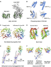
Representation of many of the protein folds discussed in this review. (A) The jelly roll fold. The structures of the major coat/capsid protein (MCP) of bacteriophage PM2 (PDB no. 2VVF) and human adenovirus 5 (1P30) are shown. The two four-stranded β-sheets that form the double jelly roll fold are colored green for clarity. (B) The PCTD. The structures of PCTD from paramyxovirus (measles virus, MeV) (1OKS), rhabdoviruses (lettuce necrotic yellows virus, LNYV; and rabies virus, RAV) (3T4R and 1VYI), and filovirus (Ebola virus, EBOV) (3FKE) are shown. The α-helical core for each structure is colored blue, as determined by Martinez et al. (2013). (C) The six-bladed β-propeller fold. Structures of the fungal Aleuria aurantia lectin (1OFZ) and the influenza A virus N4 neuraminidase (2HTV) are shown and are colored as a rainbow from the N- (blue) to C-terminus (red). Each of the six ‘blades’ of the β-propeller is labeled accordingly (β1–β6). (D) The Bcl-2 fold. Structures of the human Bcl-2 protein (1G5M) and the vaccinia virus, VACV, N1 protein (2I39) are shown. The molecules are colored as in panel C. (E) The influenza A virus hemagglutinin (HA) fold. Structures of H1 (1RUZ) and H10 (4QY1) HAs, representing groups 1 and 2 HAs, respectively, are shown. The HA1 domains are colored as a rainbow from N- (blue) to C-terminus (red), while the HA2 domains are colored white. (F) The arenaviral GP1 fold. Structures of the OW LASV GP1, with and without GP2 (4ZJF and 5VK2), and the NW JUNV GP1 (5NUZ) are shown. GP1 molecules are colored as a rainbow ramped from blue (N-terminus) to red (C-terminus). GP2 is colored white for clarity. All structures are shown in cartoon representation.
Figure 2.
Figure 2.
SBPA of six-bladed β-propeller structures. (A) Homologous six-bladed β-propeller structures were identified by application of the NDV-HN (PDB no. 1E8T) into the DALI server (Holm and Sander 1993). SBPA was subsequently performed with the following structures: MeV-H, measles virus hemagglutinin (2RKC); SosV-HN, Sosuga virus HN (6SG8); HPIV3-HN, human parainfluenza virus 3 HN (1V2I); NDV-HN; PIV5-HN, parainfluenza virus 5 HN (1Z4Y); MuV-HN, mumps virus HN (5B2C); MojV-G, Mòjiāng virus glycoprotein (5NOP); GhV-G, Ghana virus G (4UF7); NiV-G, Nipah virus G (2VSM); HeV-G, Hendra virus G (2X9M); CedV-G, Cedar virus G (6THB); influenza B virus NA molecules B/Perth (3K36), B/Lyon (4CPO), B/Brisbane (4CPL); influenza A virus NA molecules A/Vietnam N1 (2HTY), A/Tokyo N2 (1IVG), A/Missouri N3 (4HZV), A/Sweden N4 (2HTV), A/Alberta N5 (3SAL), A/England N6 (1V0Z), A/ALB N7 (4QN3), A/Ukraine N8 (2HT5), A/Australia N9 (7NN9), A/Guatemala NL10 (4FVK), A/Peru NL11 (4K3Y); Salmonellatyphimurium sialidase (3SIL); Trypanosoma rangeli sialidase (1N1S); Trypanosoma cruzi trans-sialidase (1MR5); Vibrio cholerae sialidase (1KIT); Streptococcus pneumoniae NanA (2YA4); Clostridium perfringens NanI (2VK5); S. pneumoniae NanC (4YZL); S. pneumoniae NanB (2VW0); Ruminococcus gnavus intramolecular trans-sialidase (4X47); Macrobdella decora intramolecular trans-sialidase (1SLL); Homo sapien Neu2 (1SNT); Micromonospora viridifaciens sialidase (1EUT); Aspergillus fumigatus sialidase (2XCY); Bacteroides thetaiotaomicron sialidase (4BBW); Pseudomonas aeruginosa pseudaminidase (2W38); and bacteriophage K1F endosialidase (1V0F). An evolutionary distance matrix was calculated by SHP (Stuart et al. 1979) using pairwise structural superimposition of the six-bladed β-propeller structures and an unrooted tree was plotted with PHYLIP (Felsenstein 1989). Detailed views for SBPA of paramyxovirus attachment glycoproteins (yellow circle) and influenza virus neuraminidases (green circle) are presented in panels (B) and (C), respectively. (B and C) The β-propeller structures are shown in surface representation and colored white with the receptor-binding site colored red. β-propeller structures known to bind sialic acid are highlighted with a light blue background, while β-propeller structures that bind proteinaceous receptors are highlighted with a light red background. Note, the RBP of SosV is annotated as an HN glycoprotein, however, the receptor is currently unknown (Bowden et al. 2001; Chua et al. 2002; Johnson et al. 2019; Stelfox and Bowden 2019). Calculated evolutionary distances are indicated beside the branches.
Figure 3.
Figure 3.
SBPA of the influenza A virus hemagglutinin (HA). Structures of influenza A virus HAs used were as follows: A/Brevig mission H1 (1RUZ), A/Japan H2 (2WRD), A/Finland H3 (2YP2), A/Czechoslovakia H4 (5XL5), A/Vietnam H5 (2FK0), A/New York H6 (4WSR), A/Italy H7 (1TI8), A/Hong Kong H9 (1JSD), A/Jiangxi-Donghu H10 (4QY1), A/Maryland H13 (4KPQ), A/Astrakhan H14 (3EYJ), A/Western Australia H15 (5TG8), A/Sweden H16 (4FIU), A/Guatemala HL17 (4I78), and A/Peru HL18 (4MC5). A pairwise evolutionary distance matrix was created using SHP (Stuart et al. 1979) and displayed as an unrooted phylogenetic tree using PHYLIP (Felsenstein 1989). The HA structures are shown in cartoon representation. HA1 domains for HL17 and HL18 are colored as a rainbow from the N- (blue) to C-terminus (red). HA1 domains for other HAs are colored white, while HA2 domains are colored dark gray. Branches indicating groups 1 and 2 influenza A virus HAs are colored blue and green, respectively. Calculated evolutionary distances derived from this analysis are indicated next to each branch.
Figure 4.
Figure 4.
Structure-based phylogenetic tree showing the conformations adopted by arenaviral GP1 attachment glycoproteins. Structures of available arenavirus GP1 glycoproteins used were as follows: LORV, Loei River virus (PDB no. 6HJ6); LASV, Lassa virus (4ZJF and 5VK2); MORV, Morogoro virus (5NFF); LCMV, Lymphocytic choriomeningitis virus (5INE); LUJV, Lujo virus (6GH8); WWAV, Whitewater Arroyo virus (6HJ5); MACV, Machupo virus (2WFO); and JUNV, Junín virus (5NUZ). For 5VK2, 5INE, 6GH8, and 5NUZ, all chains not comprising GP1 molecules (e.g. GP2, receptor, and antibody fragments) were removed prior to structural alignment. A pairwise evolutionary distance matrix was created using SHP (Stuart et al. 1979) and displayed as an unrooted phylogenetic tree using PHYLIP (Felsenstein 1989). GP1 structures are shown in cartoon representation and colored as a rainbow from the N- (blue) to C-terminus (red). Although the GP2 component of the GPC was not included in the structure comparison, it is shown and colored as a white cartoon for clarity. Calculated evolutionary distances derived from this analysis are indicated next to each branch.
Figure 5.
Figure 5.
SBPA of coronavirus S1-CTD glycoproteins. Structures of coronavirus S1-CTD glycoproteins used were as follows: human coronavirus HKU1 (PDB no. 5GNB); HKU4 (4QZV); HKU5 (5XGR); Middle East respiratory syndrome coronavirus, MERS-CoV (4ZPW); severe acute respiratory syndrome coronavirus, SARS-CoV (3D0I); transmissible gastroenteritis coronavirus, TGEV (4F2M); porcine respiratory coronavirus, PRCV (4F5C); human coronavirus NL63 (3KBH); human coronavirus 229E (6ATK); porcine deltacoronavirus, PDCoV (6BFU). All chains not comprising S1-CTD (e.g. receptor and antibody fragments) were removed prior to structural alignment. A pairwise evolutionary distance matrix was created using SHP (Stuart et al. 1979) and displayed as an unrooted phylogenetic tree using PHYLIP (Felsenstein 1989). Branches corresponding to alpha-, beta-, and delta-coronaviruses are colored green, blue, and brown, respectively. S1-CTD structures are shown in cartoon representation and colored as a rainbow from the N- (blue) to C-terminus (red). Although ACE2 was not included in the structure comparison, it is shown and colored here as a white cartoon. This analysis demonstrates that although SARS-CoV and NL63 S1-CTDs utilize the same receptor (Li 2005, 2008; Wu et al. 2009; Song et al. 2018), the structures classify according to genetic relationship with other coronavirus S1-CTDs, rather than receptor tropism characteristics. The viruses are color-coded according to their receptor usage: pink for dipeptidyl peptidase 4 (DPP4); orange for angiotensin converting enzyme 2 (ACE2); and gray for aminopeptidase N (APN). Calculated evolutionary distances derived from this analysis are indicated next to each branch.

References

    1. Abraham J. et al. (2009) ‘Host-Species Transferrin Receptor 1 Orthologs Are Cellular Receptors for Nonpathogenic New World Clade B Arenaviruses’, PLoS Pathogens, 5: e1000358. - PMC - PubMed
    1. Abraham J. et al. (2010) ‘Structural Basis for Receptor Recognition by New World Hemorrhagic Fever Arenaviruses’, Nature Structural & Molecular Biology, 17: 438–44. - PMC - PubMed
    1. Abrescia N. G. et al. (2008) ‘Insights into Virus Evolution and Membrane Biogenesis from the Structure of the Marine Lipid-Containing Bacteriophage PM2’, Molecular Cell, 31: 749–61. - PubMed
    1. Abrescia N. G. et al. (2012) ‘Structure Unifies the Viral Universe’, Annual Review of Biochemistry, 81: 795–822. - PubMed
    1. Ader-Ebert N. et al. (2015) ‘Sequential Conformational Changes in the Morbillivirus Attachment Protein Initiate the Membrane Fusion Process’, PLoS Pathogens, 11: e1004880. - PMC - PubMed