

Integrating AI into radiology workflow: levels of research, production, and feedback maturity
- PMID: 32064302
- PMCID: PMC7012173
- DOI: 10.1117/1.JMI.7.1.016502
Integrating AI into radiology workflow: levels of research, production, and feedback maturity
Abstract
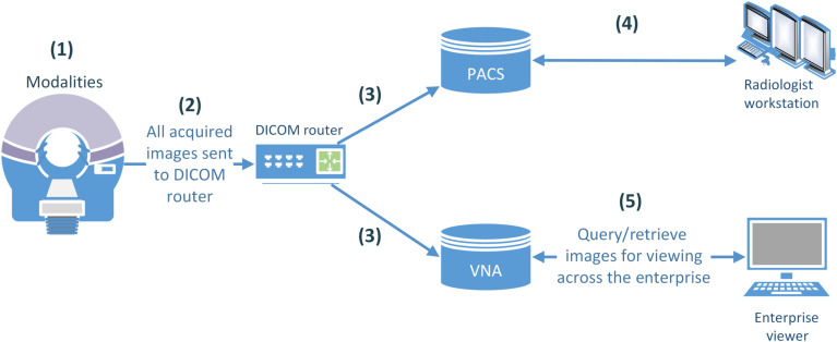
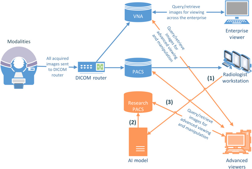
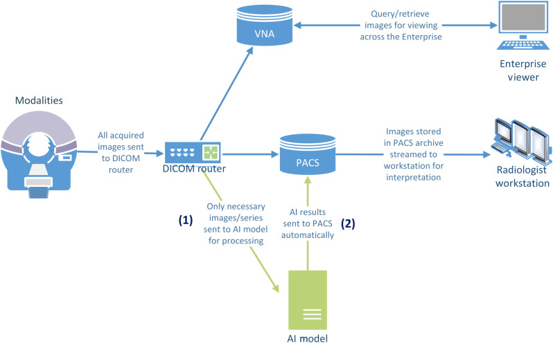
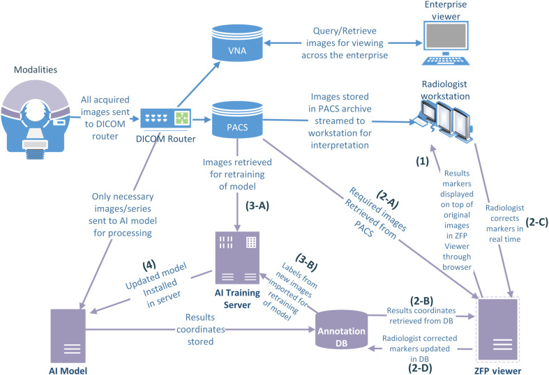
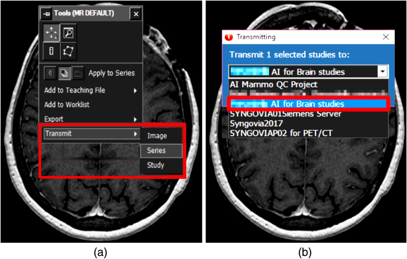
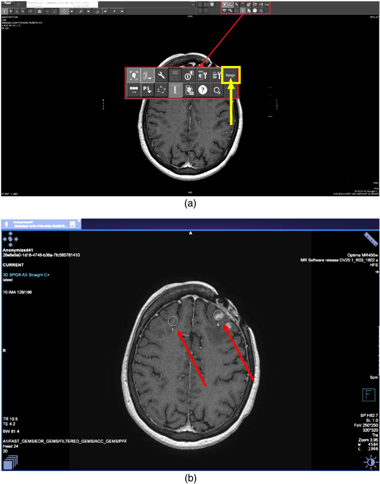
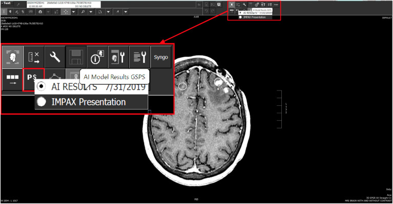
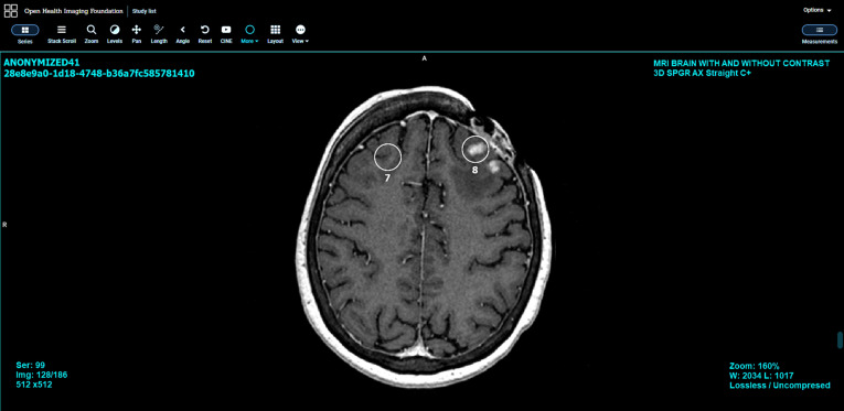
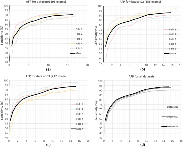
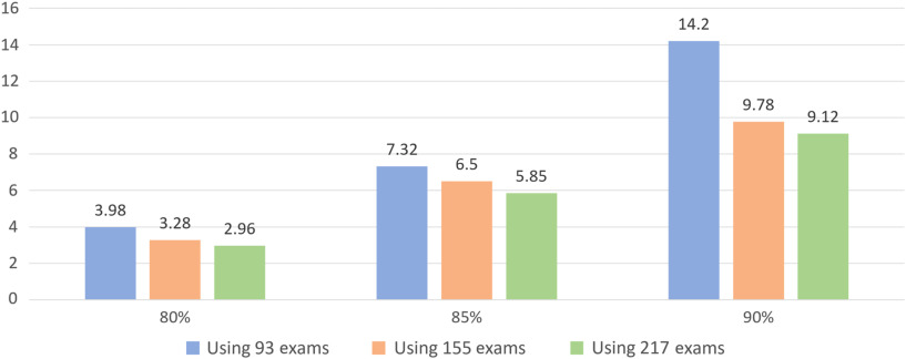
We present a roadmap for integrating artificial intelligence (AI)-based image analysis algorithms into existing radiology workflows such that (1) radiologists can significantly benefit from enhanced automation in various imaging tasks due to AI, and (2) radiologists' feedback is utilized to further improve the AI application. This is achieved by establishing three maturity levels where (1) research enables the visualization of AI-based results/annotations by radiologists without generating new patient records; (2) production allows the AI-based system to generate results stored in an institution's picture-archiving and communication system; and (3) feedback equips radiologists with tools for editing the AI inference results for periodic retraining of the deployed AI systems, thereby allowing continuous organic improvement of AI-based radiology-workflow solutions. A case study (i.e., detection of brain metastases with T1-weighted contrast-enhanced three-dimensional MRI) illustrates the deployment details of a particular AI-based application according to the aforementioned maturity levels. It is shown that the given AI application significantly improves with feedback coming from radiologists; the number of incorrectly detected brain metastases (false positives) decreases from 14.2 to 9.12 per patient with the number of subsequently annotated datasets increasing from 93 to 217 as a result of radiologist adjudication.
Keywords: AI-based image analysis; digital imaging and communications in medicine; picture archiving and communication system; radiology workflow.
© The Authors. Published by SPIE under a Creative Commons Attribution 4.0 Unported License. Distribution or reproduction of this work in whole or in part requires full attribution of the original publication, including its DOI.
Figures
References
-
- Wyawahare M. V, et al. , “Image registration techniques: an overview,” Int. J. Signal Process. Image Process. Pattern Recognit. 2(3), 11–28 (2009).
-
- Deepa S. N., Devi B. A., “A survey on artificial intelligence approaches for medical image classification,” Indian J. Sci. Technol. 4(11), 1583–1595 (2011). 10.17485/ijst/2011/v4i11/30291 - DOI
LinkOut - more resources
Full Text Sources
Other Literature Sources
