. 2020 Jan 31;6(5):eaax2659.
doi: 10.1126/sciadv.aax2659. eCollection 2020 Jan.

Single-cell analysis reveals different age-related somatic mutation profiles between stem and differentiated cells in human liver

Affiliations

Single-cell analysis reveals different age-related somatic mutation profiles between stem and differentiated cells in human liver

K Brazhnik et al. Sci Adv. .

Erratum in

Abstract

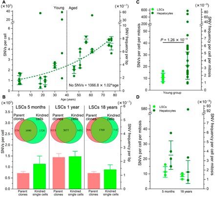
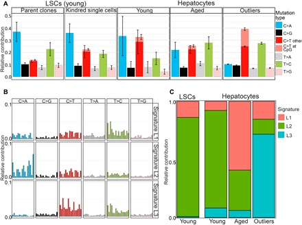
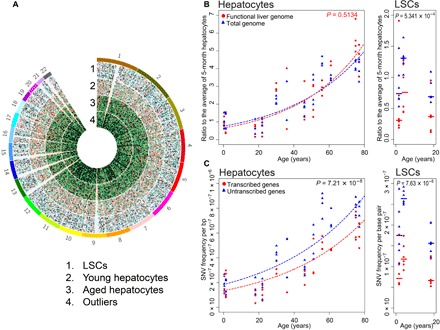
Accumulating somatic mutations have been implicated in age-related cellular degeneration and death. Because of their random nature and low abundance, somatic mutations are difficult to detect except in single cells or clonal cell lineages. Here, we show that in single hepatocytes from human liver, an organ exposed to high levels of genotoxic stress, somatic mutation frequencies are high and increase substantially with age. Considerably lower mutation frequencies were observed in liver stem cells (LSCs) and organoids derived from them. Mutational spectra in hepatocytes showed signatures of oxidative stress that were different in old age and in LSCs. A considerable number of mutations were found in functional parts of the liver genome, suggesting that somatic mutagenesis could causally contribute to the age-related functional decline and increased incidence of disease of human liver. These results underscore the importance of stem cells in maintaining genome sequence integrity in aging somatic tissues.


Figures

Fig. 1
Fig. 1. SNV levels in normal human liver cells.
(A) SNV levels in individual differentiated hepatocytes. The y axis on the left indicates the number of mutations per cell, and the y axis on the right indicates mutation frequency per base pair. The median values with SDs among four cells of each subject are indicated. Data indicate an exponential increase in mutation frequency with donor age (R = 0.892, P = 1.16 × 10⁻⁶). bp, base pair. (B) SNV levels in LSC-derived parent clones (red) and their kindred cells (light green) from three young donors. The Venn diagrams indicate the fraction of SNVs detected in the parent clones (collectively for each individual; n = 3) that were also detected in the kindred LSCs. The bars indicate the median mutation frequencies in clones (red) and kindred single cells (light green). (C) Comparison of SNV levels in differentiated hepatocytes (dark green dots; n = 24 from six donors) and LSCs (light green; n = 10 from three donors), all within the young donor group ≤36 years. Mutation frequencies were corrected for the estimated number of cell divisions. (D) SNV levels in LSCs and differentiated hepatocytes from the same participants, corrected for the estimated number of cell divisions.
Fig. 2
Fig. 2. Mutational spectra in normal human liver cells.
(A) Relative contribution of the indicated six mutation types to the point mutation spectrum for the five indicated liver sample groups. Data are represented as the mean relative contribution of each mutation type in sample groups of young and aged differentiated hepatocytes (21 cells from six donors ≤36 years, and 23 cells from six donors ≥46 years), adult LSC-derived parent clones and their kindred single cells separately, and a group of outlier cells (n = 4). (B) Three mutational signatures (L1, L2, and L3) were de novo identified by non-negative matrix factorization analysis from the somatic mutations in the different groups in (A). (C) Contributions of signatures L1, L2, and L3 to all SNVs in young and aged hepatocytes, young LSCs, and outlier cells.
Fig. 3
Fig. 3. SNV distributions across total and functional genome in human liver.
(A) Circos diagram of genomic SNV distribution in four groups: pooled LSCs, young and aged hepatocytes, and outlier cells. (B) SNV levels in the functional genome and genome overall in differentiated hepatocytes (left) and in LSCs (right) as a function of age. Each data point represents the ratio of the number of mutations per cell to the median number of mutations of the four cells from the 5-month-old subject. Mutations in the functional genome are shown in red and those in the genome overall in blue. (C) Mutation frequency per base pair in the transcribed part of the liver genome (red) and the nontranscribed part (blue) in differentiated hepatocytes (left) and LSCs (right) as a function of age.

References

    1. Maslov A. Y., Vijg J., Genome instability, cancer and aging. Biochim. Biophys. Acta 1790, 963–969 (2009). - PMC - PubMed
    1. Vijg J., Somatic mutations, genome mosaicism, cancer and aging. Curr. Opin. Genet. Dev. 26, 141–149 (2014). - PMC - PubMed
    1. Iourov I. Y., Vorsanova S. G., Yurov Y. B., Chromosomal mosaicism goes global. Mol. Cytogenet. 1, 26 (2008). - PMC - PubMed
    1. Zhang L., Vijg J., Somatic mutagenesis in mammals and its implications for human disease and aging. Annu. Rev. Genet. 52, 397–419 (2018). - PMC - PubMed
    1. Dollé M. E., Snyder W. K., Dunson D. B., Vijg J., Mutational fingerprints of aging. Nucleic Acids Res. 30, 545–549 (2002). - PMC - PubMed

Publication types