. 2020 Jan 29;6(5):eaax9318.
doi: 10.1126/sciadv.aax9318. eCollection 2020 Jan.

Modified cyclodextrins as broad-spectrum antivirals

Affiliations

Modified cyclodextrins as broad-spectrum antivirals

Samuel T Jones et al. Sci Adv. .

Abstract

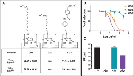
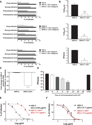
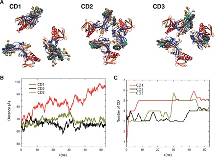
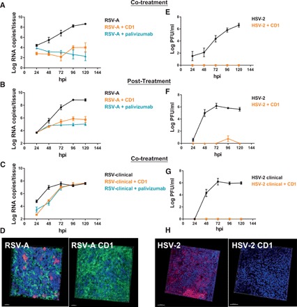
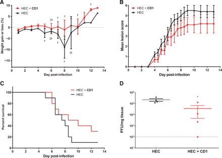
Viral infections kill millions of people and new antivirals are needed. Nontoxic drugs that irreversibly inhibit viruses (virucidal) are postulated to be ideal. Unfortunately, all virucidal molecules described to date are cytotoxic. We recently developed nontoxic, broad-spectrum virucidal gold nanoparticles. Here, we develop further the concept and describe cyclodextrins, modified with mercaptoundecane sulfonic acids, to mimic heparan sulfates and to provide the key nontoxic virucidal action. We show that the resulting macromolecules are broad-spectrum, biocompatible, and virucidal at micromolar concentrations in vitro against many viruses [including herpes simplex virus (HSV), respiratory syncytial virus (RSV), dengue virus, and Zika virus]. They are effective ex vivo against both laboratory and clinical strains of RSV and HSV-2 in respiratory and vaginal tissue culture models, respectively. Additionally, they are effective when administrated in mice before intravaginal HSV-2 inoculation. Lastly, they pass a mutation resistance test that the currently available anti-HSV drug (acyclovir) fails.


Figures

Fig. 1
Fig. 1. Structures and virucidal data for modified CDs.
(A) Structures of modified CDs and relative effective concentrations of inhibition of HSV-2 growth. (B) Dose-response assay in Vero cells. Serial dilutions of CD1, CD2, and CD3 were incubated for 1 hour at 37°C with HSV-2 and then added on cells for 2 hours at 37°C. Subsequently, cells were washed and overlaid with medium containing methylcellulose. Plaques were counted 24 hpi. Percentages of infections were calculated by comparing the number of plaques in treated and untreated wells. (C) Virucidal assays: HSV-2 was incubated with media or CDs (CD1, CD2, and CD3) at 300 μg/ml and then serially diluted on Vero cells to a negligible concentration of compound. Results are shown as the mean and SEM of three (for CD1) and two (for all other compounds) independent experiments. UT, untreated; n.a., not assessable.
Fig. 2
Fig. 2. Mechanism of action of CD1.
(A and B) CD1 was tested against RSV-A (top), DENV-2 (middle), and HSV-2 (bottom). (A) CD1 was either preincubated for 1 hour with the virus before addition on cells (Pretreatment virus), directly added with viruses on cells (During infection), added for 1 hour on cells and then washed out before infection (Pretreatment cells), or finally added after removal of the virus (Post-infection). (B) Virucidal assay: Viruses (in panels as above) were incubated with CD1 (30 μg) for 1 hour and then serially diluted on cells. (C) DNA exposure assay: HSV-2 was incubated in the presence or absence of CD1 (30 μg) for 1 hour at 37°C and then incubated for 30 min with Turbo DNase or only buffer and subsequently subjected to qPCR. Results are expressed in fold change of DNase treated versus untreated. (D) Time course of virucidal activity: HSV-2 and CD1 (30 μg) were incubated for different time periods and then serially diluted on Vero cells. (E and F) Drug resistance assay: HSV-2 passaged eight times in the presence of increasing concentrations of (E) acyclovir or (F) CD1, or no inhibitory compounds were subjected to a dose-response assay. The percentages of infection were calculated by comparing the number of plaques in treated and untreated wells. Results are mean and SEM of at least two independent experiments. *P < 0.05, **P < 0.01, ***P < 0.005.
Fig. 3
Fig. 3. MD simulations.
Interactions of CDs with gB proteins. (A) Representative snapshots of the modified CDs interacting with HSV-2 gB proteins after 50 ns of MD simulations. (B) Average distance between gB fusion loops, defined as the instantaneous average of the three distances labeled in fig. S9, over time. (C) Number of CDs interacting with gB proteins over time.
Fig. 4
Fig. 4. Ex vivo experiments.
(A) Co-treatment of respiratory tissues with RSV-A and CD1 (10 μg). (B) Respiratory tissues infected with RSV-A and treated with CD1 (6 μg) 24 hpi (Post-treatment). (C) Co-treatment of respiratory tissues with a clinical strain of RSV-A and CD1 (10 μg). (D) Respiratory tissues infected with RSV-A or cotreated with RSV-A and CD1 were subjected to immunostaining at 7 days post-infection (dpi). Green, ciliated cells (tubulin); red, infected cells; blue, nuclei. Scale bars, 20 μm. (E) Co-treatment of vaginal tissues with HSV-2 and CD1 (50 μg). (F) Vaginal tissues infected with HSV-2 and treated with CD1 (30 μg) 8 hpi post-treatment. (G) Co-treatment of vaginal tissues with a clinical strain of HSV-2 and CD1 (50 μg). Results are mean and SEM of two independent experiments. (H) Vaginal tissues infected with HSV-2 or cotreated with HSV-2 and CD1 were subjected to immunostaining at 7 dpi. Red, infected cells; blue, nuclei. Scale bars, 100 μm.
Fig. 5
Fig. 5. Intravaginal HSV-2 infection of mice.
(A) Percentage of weight changes, (B) mean lesion scores, (C) survival rates, and (D) viral titers in vaginal mucosa of BALB/c mice pretreated with 15 μl of HEC gel or 0.03 mg of CD1 per mouse in HEC gel (15 μl). Five minutes later, mice were infected intravaginally with 1.2 × 10⁵ PFU of HSV-2 strain 333 in a 5-μl volume. Vaginal mucosa was taken on day 3 after infection in a subset of mice, and viral titers were determined by plaque assay on Vero cells. Results represent the mean of 10 mice per group (A to C) or the mean and SEM of 4 mice per group (D). †, mouse death. *P < 0.05.

References

    1. De Clercq E., Strategies in the design of antiviral drugs. Nat. Rev. Drug Discov. 1, 13–25 (2002). - PubMed
    1. McCormack S., Ramjee G., Kamali A., Rees H., Crook A. M., Gafos M., Jentsch U., Pool R., Chisembele M., Kapiga S., Mutemwa R., Vallely A., Palanee T., Sookrajh Y., Lacey C. J., Darbyshire J., Grosskurth H., Profy A., Nunn A., Hayes R., Weber J., PRO2000 vaginal gel for prevention of hiv-1 infection (microbicides development programme 301): A phase 3, randomised, double-blind, parallel-group trial. Lancet 376, 1329–1337 (2010). - PMC - PubMed
    1. Pirrone V., Wigdahl B., Krebs F. C., The rise and fall of polyanionic inhibitors of the human immunodeficiency virus type 1. Antivir. Res. 90, 168–182 (2011). - PubMed
    1. Van Damme L., Govinden R., Mirembe F. M., Guédou F., Solomon S., Becker M. L., Pradeep B., Krishnan A., Alary M., Pande B., Ramjee G., Deese J., Crucitti T., Taylor D.; CS Study Group , Lack of effectiveness of cellulose sulfate gel for the prevention of vaginal hiv transmission. N. Engl. J. Med. 359, 463–472 (2008). - PubMed
    1. Rusnati M., Vicenzi E., Donalisio M., Oreste P., Landolfo S., Lembo D., Sulfated K5 Escherichia coli polysaccharide derivatives: A novel class of candidate antiviral microbicides. Pharmacol. Ther. 123, 310–322 (2009). - PubMed

Publication types

MeSH terms