. 2020 Mar;34(2):549-573.
doi: 10.1111/jvim.15725. Epub 2020 Feb 17.

ACVIM consensus statement guidelines for the diagnosis, classification, treatment, and monitoring of pulmonary hypertension in dogs

Affiliations

ACVIM consensus statement guidelines for the diagnosis, classification, treatment, and monitoring of pulmonary hypertension in dogs

Carol Reinero et al. J Vet Intern Med. 2020 Mar.

Abstract

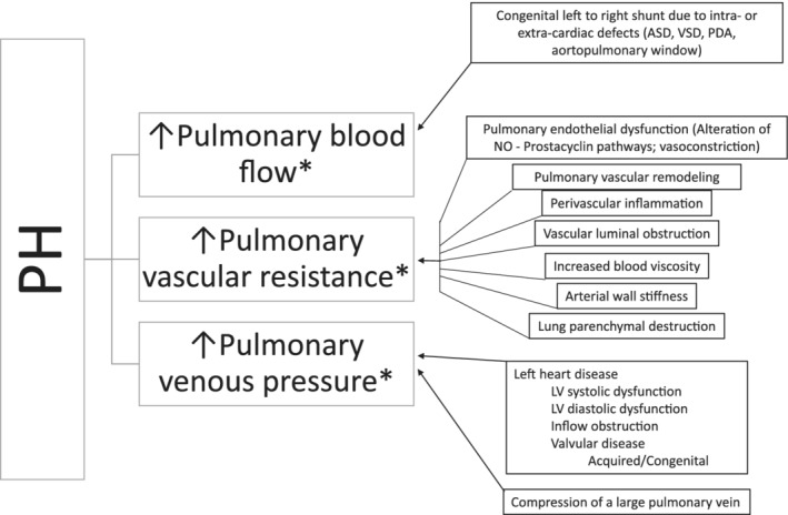
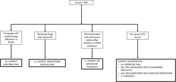
Pulmonary hypertension (PH), defined by increased pressure within the pulmonary vasculature, is a hemodynamic and pathophysiologic state present in a wide variety of cardiovascular, respiratory, and systemic diseases. The purpose of this consensus statement is to provide a multidisciplinary approach to guidelines for the diagnosis, classification, treatment, and monitoring of PH in dogs. Comprehensive evaluation including consideration of signalment, clinical signs, echocardiographic parameters, and results of other diagnostic tests supports the diagnosis of PH and allows identification of associated underlying conditions. Dogs with PH can be classified into the following 6 groups: group 1, pulmonary arterial hypertension; group 2, left heart disease; group 3, respiratory disease/hypoxia; group 4, pulmonary emboli/pulmonary thrombi/pulmonary thromboemboli; group 5, parasitic disease (Dirofilaria and Angiostrongylus); and group 6, disorders that are multifactorial or with unclear mechanisms. The approach to treatment of PH focuses on strategies to decrease the risk of progression, complications, or both, recommendations to target underlying diseases or factors contributing to PH, and PH-specific treatments. Dogs with PH should be monitored for improvement, static condition, or progression, and any identified underlying disorder should be addressed and monitored simultaneously.

Keywords: echocardiography; pulmonary arterial hypertension; respiratory disease; tricuspid regurgitation velocity.


Conflict of interest statement

Carol Reinero received donation of medication (tadalafil) for an intramurally funded clinical trial comparing efficacy of sildenafil and tadalafil in dogs with pulmonary hypertension (2017‐2018). Brian Scansen has received speaking fees and travel expense reimbursement from Boehringer Ingelheim Animal Health. All other authors have no declared conflicts of interest.

Figures

Figure 1
Figure 1
Development of pulmonary hypertension, defined as abnormally increased pressure within the pulmonary vasculature, results from increased pulmonary blood flow, increased pulmonary vascular resistance, increased pulmonary venous pressure, or some combination thereof. Normal pulmonary vasculature is comprised of thin‐walled arteries, veins and capillaries; a low pressure, low vascular resistance, and high capacitance system. In homeostasis, blood ejected from the right ventricle (RV) into the pulmonary trunk is directed to the right and left pulmonary arteries. The pulmonary arterial pressure remains low in homeostatic conditions by the dense network of pulmonary capillaries that can accommodate rapid transit of large volume of blood arriving from the pulmonary arteries. Following efficient gas exchange at the alveolar‐capillary interface, oxygenated blood is then collected by the pulmonary venules that unite to form the veins, which eventually open into the left atrium. Upon complex interactions of genetic and environmental factors, most of which are still poorly understood in dogs, homeostasis of pulmonary circulation can be disturbed. These disturbances (summarized in boxes) can lead to an excessive increase in pulmonary blood flow, increased pulmonary vascular resistance, or increased pulmonary venous pressure. Additionally, there is interplay between these factors*: increased pulmonary blood flow or increased pulmonary venous pressure can lead to increased pulmonary vascular resistance due to pulmonary arterial vasoconstriction, pulmonary vascular disease/remodeling, or both. When the average pulmonary arterial pressure increases above a certain threshold (25 mm Hg is commonly used in humans), PH results. With sustained PH, the RV has to work harder against increases in pulmonary pressures to move blood through the pulmonary vasculature. As a consequence, the RV undergoes structural alterations. Over time, the progressive increase in RV workload can ultimately lead to RV dysfunction and failure, resulting in heart failure (ascites), low output signs, and death. ASD, atrial septal defect; EDD, endothelial‐dependent dilatation; LV, left ventricle; NO, nitric oxide; PDA, patent ductus arteriosus; PH, pulmonary hypertension; VSD, ventricular septal defect
Figure 2
Figure 2
Example measurements of tricuspid regurgitation velocity (TRV) spectra acquired using continuous wave Doppler. A, The solid arrow shows the recommended measurement of TRV. The dense outer edge of the velocity profile (brighter signal) is measured and measurement sof the fine linear signals has been avoided. The dotted arrow represents a measurement that includes the fine linear signals or “feathered edge,” which likely overestimates TRV. B, A TRV signal of good quality is shown. Notice the envelope is fully visible, the signal is not overgained (helps to avoid fine linear signals), the sweep speed is increased, and the scale along the vertical axis is nearly filled by the TRV signal. C, Shows 3 TRV signals, 1 of moderate quality (*) that is not completely filled in but can be measured by extrapolation and 2 others of poor quality (#) that are unreliable and should not be measured
Figure 3
Figure 3
Algorithm demonstrating the overall diagnostic approach to the 6 groups of pulmonary hypertension in which echocardiography performed early in the clinical evaluation identifies intermediate or high probability PH. In addition to determining an intermediate or high probability of PH, echocardiography can also be used to support, confirm, or refute pathology in group 1d1, group 2, group 4, and group 5. The order in which diagnostic algorithms should be consulted are group 3 (Figure 4) and group 4 (Figure 5) with group 5 potentially being identified on this initial algorithm or within the group 3 algorithm. The group 1 algorithm (Figure 7) generally used after ruling out disorders in groups 2‐6. Critical to appropriate use of the diagnostic algorithms is the understanding that dogs frequently have greater than 1 type of pathology contributing to PH either across groups (eg, a dog with MMVD with interstitial lung disease is encompassed in groups 2 and 3, respectively) or within a group (eg, a dog with tracheal collapse and fibrotic lung disease both fall within group 3). Clinical evaluation must drive the diagnostic approach and make sense in context of localizing disease and pursuit of comorbid conditions. For example, a small breed dog with left‐sided heart failure that has inspiratory stridor in addition to rapid, shallow breathing should not have the diagnostic algorithm terminated after diagnosis of group 2c1a disease; instead, further evaluation for an upper airway defect such as extrathoracic tracheal collapse should be pursued. aThoracic radiographs are frequently obtained before echocardiography and may provide additional findings supportive of underlying PH etiology. bEvidence of an in situ PT in the main pulmonary artery may be noted on echocardiographic examination. LHD, left‐sided heart disease; PH, pulmonary hypertension, PT, pulmonary thrombus
Figure 4
Figure 4
Diagnostic algorithm for discrimination of group 3 respiratory disease/hypoxia in dogs. Proper interpretation relies on confirmation that the comprehensive clinical picture can be explained solely by the “final diagnosis” (bold boxes); otherwise, continue to evaluate for PH in other subcategories of group 3 and in groups 1, 4, 5, and 6. BAL, bronchoalveolar lavage; CT, computed tomography; FNA, fine‐needle aspiration; HW, heartworm; I/E, paired inspiratory/expiratory series; MSB, mainstem bronchial; OSA, obstructive sleep apnea; PAH, pulmonary arterial hypertension; PH, pulmonary hypertension; PTE, pulmonary thromboembolism
Figure 5
Figure 5
Diagnostic algorithm for discrimination of group 4 pulmonary emboli/thrombi/thromboemboli in dogs. Proper interpretation relies on confirmation that the comprehensive clinical picture can be explained solely by the “final diagnosis” of PE/PT/PTE; otherwise, continue to evaluate for PH in groups 1 and 6. aRisk factors include but are not limited to hypercoagulability (eg, CBC, serum biochemical profile, UA, UP:C, TEG, D‐dimers), pulmonary arterial mass, endothelial injury (eg, IV catheter, polytrauma with immobility), and evidence of air or fat emboli. bIdeally triphasic angiography is recommended. cVentilation‐perfusion scans using nuclear scintigraphy can also be used to document PE/PT/PTE but are not commonly performed. CT, computed tomography; PH, pulmonary hypertension, UA, urinalysis; UP:C, urine protein:creatitine ratio; TEG, thromboelastography
Figure 6
Figure 6
Diagnostic algorithm for determination of group 6 (multifactorial and/or unclear mechanisms) in dogs. Confirm that the comprehensive clinical picture can be explained solely by the “final diagnosis” (bold boxes). Otherwise, consider hematologic, systemic, and metabolic disorders of unclear mechanism that have been identified in humans with PH148 and if not present or likely to be causative of PH, continue to evaluate for PH in group 1. Each disease identified must be addressed in the overall treatment plan. aEach will need to be addressed independently when considering optimal treatment. CT, computed tomography; HW, heartworm; LHD, left heart disease; PA, pulmonary artery; PH, pulmonary hypertension; PE/PT/PTE, pulmonary emboli/thrombi/thromboemboli
Figure 7
Figure 7
Diagnostic algorithm for discrimination of group 1 pulmonary arterial hypertension in dogs. The approach to group 1 disorders generally requires ruling out groups 2‐6 disorders first. Importantly, histologic changes associated with the pulmonary vasculature in group 1a‐c are not pathognomonic and can occur secondary to primary cardiac and respiratory disease. Histopathology can provide definitive diagnosis for group 1d2, 1d3, and 1′ disorders. aExtrapolated from humans; not definitively proven for canine PVOD/PCH. bConfirmed on histopathology. PAH, pulmonary arterial hypertension; PH, pulmonary hypertension, PDE5, phosphodiesterase 5; PVOD/PCH, pulmonary veno‐occlusive disease/pulmonary capillary hemangiomatosis

Similar articles

Cited by

References

    1. Thangaratinam S, Redman CWE. The Delphi technique. Obstet Gynaecol. 2005;7:120‐125.
    1. Hsu CC, Sandford BA. The Delphi technique: making sense of consensus. Pract Assess Res Eval. 2007;12:1‐7.
    1. Hoeper MM, Bogaard HJ, Condliffe R, et al. Definitions and diagnosis of pulmonary hypertension. J Am Coll Cardiol. 2013;62:D42‐D50. - PubMed
    1. Simonneau G, Montani D, Celermajer DS, et al. Haemodynamic definitions and updated clinical classification of pulmonary hypertension. Eur Respir J. 2019;53:pii:13993003.01913‐2018. 10.1183/13993003.01913-2018. - DOI - PMC - PubMed
    1. Vachiery JL, Adir Y, Barbera JA, et al. Pulmonary hypertension due to left heart diseases. J Am Coll Cardiol. 2013;62:D100‐D108. - PubMed

Publication types

MeSH terms