

A critical role of the KCa 3.1 channel in mechanical stretch-induced proliferation of rat bone marrow-derived mesenchymal stem cells
- PMID: 32065503
- PMCID: PMC7131943
- DOI: 10.1111/jcmm.15014
A critical role of the KCa 3.1 channel in mechanical stretch-induced proliferation of rat bone marrow-derived mesenchymal stem cells
Abstract
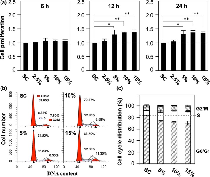
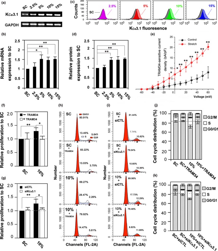
Mechanical stimulation is an important factor regulating mesenchymal stem cell (MSC) functions such as proliferation. The Ca2+ -activated K+ channel, KCa 3.1, is critically engaged in MSC proliferation but its role in mechanical regulation of MSC proliferation remains unknown. Here, we examined the KCa 3.1 channel expression and its role in rat bone marrow-derived MSC (BMSC) proliferation in response to mechanical stretch. Application of mechanical stretch stimulated BMSC proliferation via promoting cell cycle progression. Such mechanical stimulation up-regulated the KCa 3.1 channel expression and pharmacological or genetic inhibition of the KCa 3.1 channel strongly suppressed stretch-induced increase in cell proliferation and cell cycle progression. These results support that the KCa 3.1 channel plays an important role in transducing mechanical forces to MSC proliferation. Our finding provides new mechanistic insights into how mechanical stimuli regulate MSC proliferation and also a viable bioengineering approach to improve MSC proliferation.
Keywords: KCa3.1 channel; bone marrow-derived mesenchymal stem cells; cell proliferation; mechanical stretch.
© 2020 The Authors. Journal of Cellular and Molecular Medicine published by Foundation for Cellular and Molecular Medicine and John Wiley & Sons Ltd.
Conflict of interest statement
The authors confirm that there are no conflicts of interest.
Figures
Similar articles
-
Cell cycle-dependent expression of potassium channels and cell proliferation in rat mesenchymal stem cells from bone marrow.Cell Prolif. 2007 Oct;40(5):656-70. doi: 10.1111/j.1365-2184.2007.00458.x. Cell Prolif. 2007. PMID: 17877608 Free PMC article.
-
Calcium-dependent potassium channels control proliferation of cardiac progenitor cells and bone marrow-derived mesenchymal stem cells.J Physiol. 2018 Jun;596(12):2359-2379. doi: 10.1113/JP275388. Epub 2018 May 5. J Physiol. 2018. PMID: 29574723 Free PMC article.
-
Functional ion channels in mouse bone marrow mesenchymal stem cells.Am J Physiol Cell Physiol. 2007 Nov;293(5):C1561-7. doi: 10.1152/ajpcell.00240.2007. Epub 2007 Aug 15. Am J Physiol Cell Physiol. 2007. PMID: 17699636
-
Mesenchymal stem cell differentiation: Control by calcium-activated potassium channels.J Cell Physiol. 2018 May;233(5):3755-3768. doi: 10.1002/jcp.26120. Epub 2017 Sep 7. J Cell Physiol. 2018. PMID: 28776687 Review.
-
Ca2+ signalling in fibroblasts and the therapeutic potential of KCa3.1 channel blockers in fibrotic diseases.Br J Pharmacol. 2020 Mar;177(5):1003-1024. doi: 10.1111/bph.14939. Epub 2020 Feb 3. Br J Pharmacol. 2020. PMID: 31758702 Free PMC article. Review.
Cited by
-
Ca2+-Activated K+ Channels in Progenitor Cells of Musculoskeletal Tissues: A Narrative Review.Int J Mol Sci. 2023 Apr 5;24(7):6796. doi: 10.3390/ijms24076796. Int J Mol Sci. 2023. PMID: 37047767 Free PMC article. Review.
-
Gli1+ Cells Residing in Bone Sutures Respond to Mechanical Force via IP3R to Mediate Osteogenesis.Stem Cells Int. 2021 Aug 12;2021:8138374. doi: 10.1155/2021/8138374. eCollection 2021. Stem Cells Int. 2021. PMID: 34434241 Free PMC article.
-
Mechanical Force Modulates Alveolar Bone Marrow Mesenchymal Cells Characteristics for Bone Remodeling during Orthodontic Tooth Movement through Lactate Production.Cells. 2022 Nov 22;11(23):3724. doi: 10.3390/cells11233724. Cells. 2022. PMID: 36496983 Free PMC article.
-
[Role of Piezo mechanosensitive ion channels in the osteoarticular system].Zhongguo Xiu Fu Chong Jian Wai Ke Za Zhi. 2024 Feb 15;38(2):240-248. doi: 10.7507/1002-1892.202310092. Zhongguo Xiu Fu Chong Jian Wai Ke Za Zhi. 2024. PMID: 38385239 Free PMC article. Review. Chinese.
-
Mechanical regulation of bone remodeling.Bone Res. 2022 Feb 18;10(1):16. doi: 10.1038/s41413-022-00190-4. Bone Res. 2022. PMID: 35181672 Free PMC article. Review.
References
-
- Pittenger MF, Mackay AM, Beck SC, et al. Multilineage potential of adult human mesenchymal stem cells. Science. 1999;284:143‐147. - PubMed
-
- Caplan AI. Adult mesenchymal stem cells for tissue engineering versus regenerative medicine. J Cell Physiol. 2007;213:341‐347. - PubMed
-
- Banfi A, Muraglia A, Dozin B, Mastrogiacomo M, Cancedda R, Quarto R. Proliferation kinetics and differentiation potential of ex vivo expanded human bone marrow stromal cells: Implications for their use in cell therapy. Exp Hematol. 2000;28:707‐715. - PubMed
-
- Park JS, Chu JS, Cheng C, Chen F, Chen D, Li S. Differential effects of equiaxial and uniaxial strain on mesenchymal stem cells. Biotechnol Bioeng. 2004;88:359‐368. - PubMed
Publication types
MeSH terms
Substances
LinkOut - more resources
Full Text Sources
Miscellaneous
