Comparative Study
. 2020 Feb 18;27(1):35.
doi: 10.1186/s12929-020-00629-y.

Treatment of rats with spinal cord injury using human bone marrow-derived stromal cells prepared by negative selection

Affiliations
Comparative Study

Treatment of rats with spinal cord injury using human bone marrow-derived stromal cells prepared by negative selection

Lorenzo Romero-Ramírez et al. J Biomed Sci. .

Abstract

Background: Spinal cord injury (SCI) is a highly debilitating pathology without curative treatment. One of the most promising disease modifying strategies consists in the implantation of stem cells to reduce inflammation and promote neural regeneration. In the present study we tested a new human bone marrow-derived stromal cell preparation (bmSC) as a therapy of SCI.

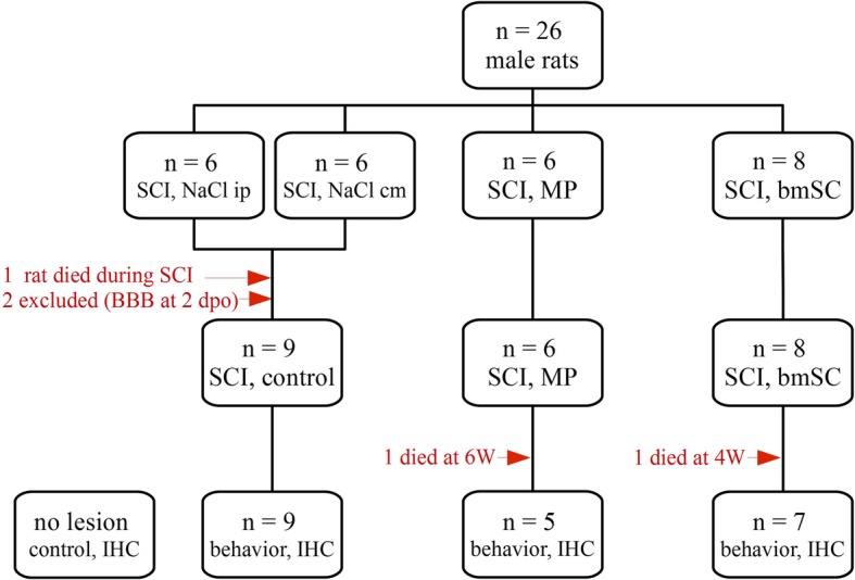
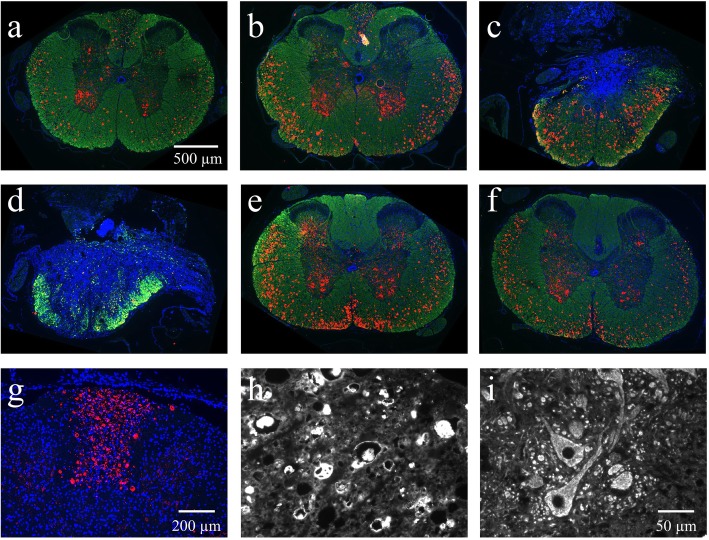
Methods: Spinal cord contusion injury was induced in adult male rats at thoracic level T9/T10 using the Infinite Horizon impactor. One hour after lesion the animals were treated with a sub-occipital injection of human bmSC into the cisterna magna. No immune suppression was used. One dose of bmSC consisted, on average, of 2.3 million non-manipulated cells in 100 μL suspension, which was processed out of fresh human bone marrow from the iliac crest of healthy volunteers. Treatment efficacy was compared with intraperitoneal injections of methylprednisolone (MP) and saline. The recovery of motor functions was assessed during a surveillance period of nine weeks. Adverse events as well as general health, weight and urodynamic functions were monitored daily. After this time, the animals were perfused, and the spinal cord tissue was investigated histologically.

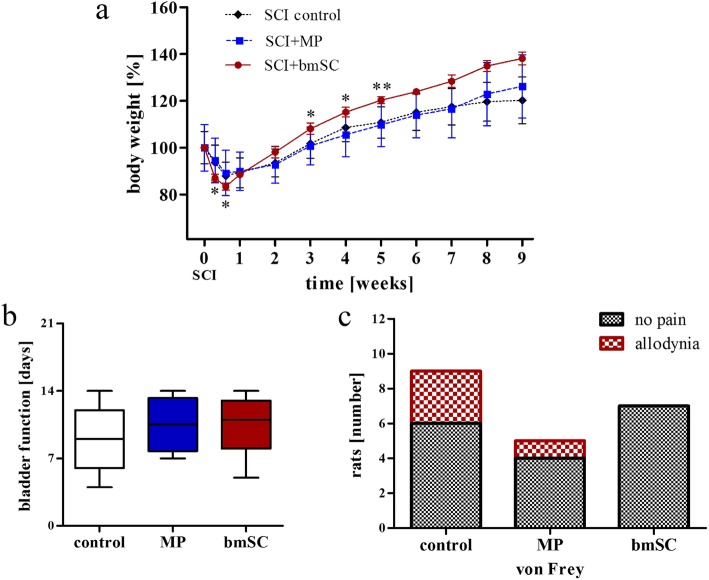
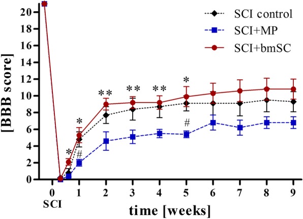
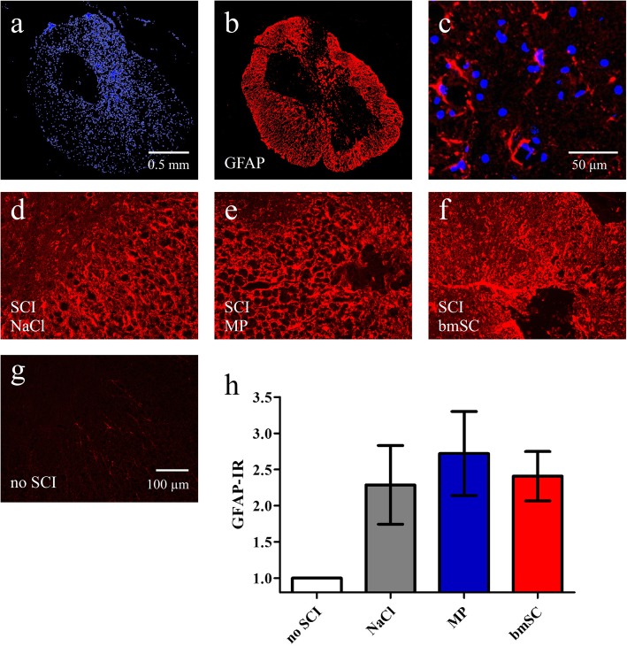
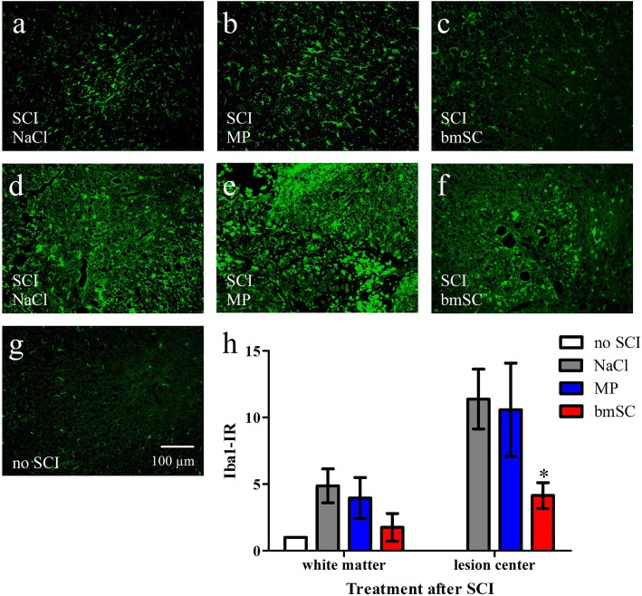
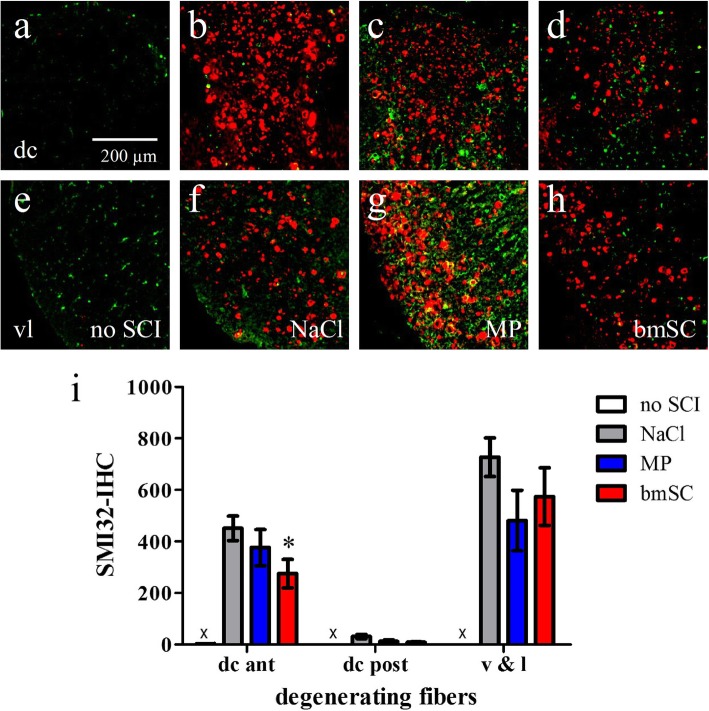
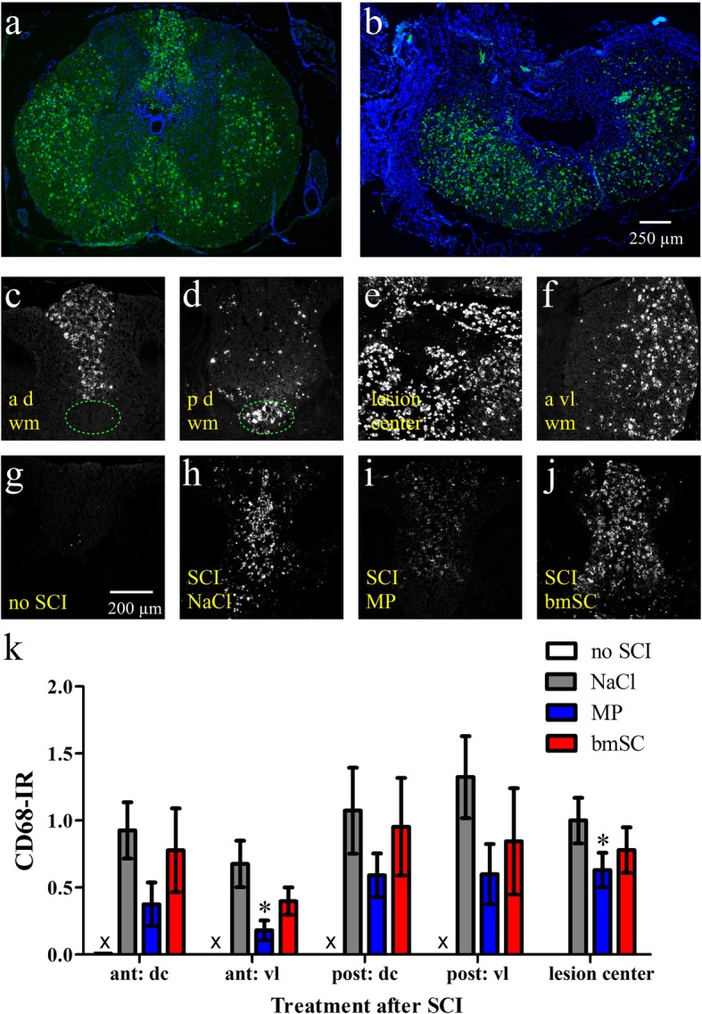
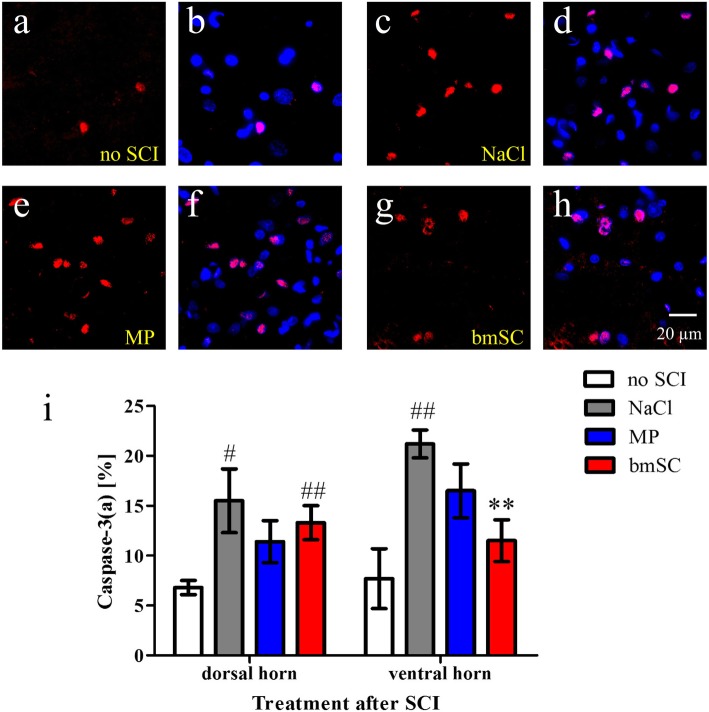
Results: Rats treated with bmSC did not reject the human implants and showed no sign of sickness behavior or neuropathic pain. Compared to MP treatment, animals displayed better recovery of their SCI-induced motor deficits. There were no significant differences in the recovery of bladder control between groups. Histological analysis at ten weeks after SCI revealed no differences in tissue sparing and astrogliosis, however, bmSC treatment was accompanied with reduced axonal degeneration in the dorsal ascending fiber tracts, lower Iba1-immunoreactivity (IR) close to the lesion site and reduced apoptosis in the ventral grey matter. Neuroinflammation, as evidenced by CD68-IR, was significantly reduced in the MP-treated group.

Conclusions: Human bmSC that were prepared by negative selection without expansion in culture have neuroprotective properties after SCI. Given the effect size on motor function, implantation in the acute phase was not sufficient to induce spinal cord repair. Due to their immune modulatory properties, allogeneic implants of bmSC can be used in combinatorial therapies of SCI.

Keywords: Bone marrow; Human; Inflammation; Rat; Spinal cord injury; Stem cells.


Conflict of interest statement

JdM and EChW are CEOs and owners of Neuroplast BV.

Figures

Fig. 1
Fig. 1
Experimental plan and treatment groups. Animals that received spinal cord contusion injury were pseudo-randomly assigned to four groups. Rats with intraperitoneal and intrathecal (cisterna magna) saline injections were planned to be evaluated as one control group unless behavioral evaluation showed statistical differences between them. One animal was lost due to bleeding during spinal cord surgery, two animals had to be excluded from the study because open field evaluation revealed an incomplete lesion (BBB at 2 dpo) and two rats died during the observation period. In the histological evaluation, treatment groups were also compared to tissue samples of non-injured animals
Fig. 2
Fig. 2
Health status after SCI. No adverse events were attributed to bmSC treatment. a Changes of body weight following SCI: Initially, stem cell-treated rats lost more body weight while at a later stage (starting at 14 dpo) they gained more weight compared to MP- and vehicle-treated animals. Data were normalized to the body weight before surgery (mean +/− SEM; two factor ANOVA, post hoc Tukey test, * p < 0.05, ** p < 0.01). b Recovery of the spontaneous micturition reflex: Displayed is the time after SCI [days] that passed until the animals no longer required manual voiding of the bladder by the experimenter (median, 25%/95% and range). There were no significant differences between treatment groups (H-test). c Testing of mechanical nociception (von Frey, reduced threshold of paw withdrawal response) at nine weeks after SCI showed no hyperalgesia/allodynia in bmSC implanted animals, while this occurred in 1/5 rats treated with MP and 3/9 rats that had received NaCl injections. Treatments following SCI are designated as: control - injections of 0.8% saline solution; MP - of methyl prednisolone; bmSC - of human bone marrow-derived stem cells
Fig. 3
Fig. 3
Treatment with bmSC and MP did not affect tissue degeneration. Lesion size and extent of tissue degeneration were evaluated in H&E-stained spinal cord sections at 10 weeks after SCI. a-f Panels show representative tissue sections 0.9 cm anterior of the lesion site (a-c) and at the lesion center (d-f); treatment groups were: a, d injection of saline; b, e methylprednisolone; and c, f human bmSC; same magnification in all photographs. g Relative tissue loss in the center of the lesion (normalized to spinal cord sections without lesion). h Anterior to posterior extension of lesion size as identified in H&E-stained spinal cord sections. Bars show means and SEM, n = 5–7 animals, differences between treatment groups were not significant
Fig. 4
Fig. 4
Injection of bmSC caused better recovery of motor function than MP treatment. Mean BBB scores (± SEM) of the three treatment groups. All rats had BBB = 21 before SCI, and the first evaluation occurred two days after surgery. As indicated in Fig. 1, only animals with BBB < 2 at 2 dpo (SCI considered as complete) were included in the evaluation. Following a two-factor ANOVA that revealed effects of treatment and time after SCI, post hoc Tukey test showed significant differences between bmSC and MP treatment (* p < 0.05, ** p < 0.01) and between NaCl and MP treatment (♯ p < 0.05)
Fig. 5
Fig. 5
Astrogliosis was not affected by bmSC and MP treatment. Evaluation of GFAP-IR in spinal cord tissue ten weeks after SCI. a-b Overview of scar formation around the lesion center in a typical example; nuclear labeling with Hoechst-33342 (a) was combined with GFAP (b) immunostaining; 5x objective, scale bar 0.5 mm in a. c Reactive astrocytes in the gey matter outside of the lesion center. d-f Higher magnification of GFAP-IR close to the lesion site in SCI rats with control treatment (d), MP injections (e), bmSC implants (f), and g in the white matter of an animal without SCI; 20x objective, images d-g with the same times of exposure, scale bar 100 μm in g. h Quantification of GFAP-IR (integrated density) near the lesion site revealed no significant differences between SCI treatment groups (t-tests, p > 0.5). Data were normalized to GFAP-IR in the white matter of rats without lesion (statistical difference not indicated); bars show means and SEM, n = 5–6 rats/group
Fig. 6
Fig. 6
Injection of bmSC reduced activation of microglia/macrophages. Evaluation of Iba1-IR in spinal cord tissue ten weeks after SCI. a-c Microglia in spinal cord white matter 0.5–0.7 cm anterior of the lesion center. d-f Microglia and macrophages in sections containing the lesion center; representative examples from rats treated with saline (a, d), MP (b, e) and bmSC (c, f). g Microglia in the white matter of an animal without SCI; 20 objective, scale bar 100 μm valid for all photographs. h Quantification of Iba1-IR in the white matter ca. 0.8 cm anterior to and within the area close to the lesion center. Here, Iba1 expression was significantly lower after bmSC treatment compared to control treatment (t-test, * p < 0.5). Data were normalized to Iba1-IR in the white matter of rats without lesion (statistical difference not indicated); bars show means and SEM, n = 5–6 rats/group
Fig. 7
Fig. 7
The presence of non-phosphorylated filaments as an indicator of axonal damage. Ten weeks after SCI immune staining with Smi32 antibody (red) was combined with myelin basic protein-IR (green) and Hoechst-33342 nuclear staining (blue). a-f Overview of transverse spinal cord sections at intervals of approximately 3.2 mm from 8 mm anterior to 8 mm posterior of the lesion site; 5x objective, scale bar in a. Note the presence of Smi32-binding in the ascending dorsal columns anterior but not posterior of the lesion site and in white matter tracts in all sections. g Non-phosphorylated neurofilament in ascending fiber tracts anterior of the site of injury, 20x objective. h-i Higher magnification of Smi32-IR in white matter (h) and motor neurons in the ventral horn (i), 40x objective, scale bar in i. No Smi32 staining was observed in the white matter of animals without SCI (see Fig. 8)
Fig. 8
Fig. 8
Treatment with bmSC reduced axonal damage in ascending fiber tracts anterior of the lesion site. Staining with Smi32 (red) was combined with Iba1 (green) in spinal cord tissue ten weeks after SCI. a-h Smi32 IR in the dorsal columns (a-d) and ventrolateral white matter (e-h) of a rat without SCI (a, e), and of SCI animals treated with saline (b, f), MP (c, g) and bmSC injections (d, h); 20 objective, scale bar in a. Note the absence of non-phosphorylated neurofilament in control samples without SCI in a and e. i Quantification of Smi32-IR in the ascending dorsal columns anterior and posterior of the lesion site (dc ant, dc post), the ventrolateral white matter (v&l) and corresponding regions without SCI (no Smi32-IR, marked x). Bars show means and SEM, n = 5–6 rats/group. Treatment with bmSC was associated with reduced Smi32-IR in the anterior dorsal columns compared to saline treatment (t-test * p < 0.05), while MP had no effect and differences in dc post and vl were not significant
Fig. 9
Fig. 9
Injections of MP mitigated SCI-induced neuroinflammation. Microglia activation and macrophage infiltration ten weeks after SCI were evaluated with immune staining of CD68 (ED1). a-b Overview of transverse spinal cord sections 0.8 cm anterior of the lesion site and at its center. CD68 (green) was combined with Hoechst-33342 nuclear staining (blue), 5x objective, scale bar in b. Note very strong CD68-IR everywhere in the white matter as well as its absence in the scar tissue (b). c-f Examples of activated microglia/macrophages in ascending fiber tracts in the dorsal columns anterior of the lesion site (c), in corticospinal tract posterior of the lesion center (d; marked with dotted ellipse in c and d), in the lesion center (e), and anterior ventrolateral white matter (f). g-j Examples of CD68-IR in dorsal columns of rats without SCI and after SCI treatments; 10x objective, scale bar in g. k Quantification of CD68-IR in the dorsal columns (dc) and ventrolateral white matter (vl) anterior and posterior of the lesion site and corresponding regions without SCI (no CD68-IR). Bars show means and SEM, n = 5 rats/group. As indicated (t-test * p < 0.05) treatment with MP was associated with reduced CD68-IR compared to saline treatment. Injections of bmSC had no significant effect
Fig. 10
Fig. 10
Injection of bmSC reduced apoptosis in the ventral horn. Ten weeks after SCI, cellular apoptosis was evaluated using activated caspase-3 as a marker. a-h Representative ROI containing apoptotic nuclei in the ventral horns of non-injured animals (a, b), after SCI/treatment with saline (c, d), with MP (e, f) and with bmSC (g, h). Immune staining of activated caspase-3 (red, all panels) was combined with Hoechst-33342 nuclear staining (blue, b, d, f, h, double exposure); 40x objective, scale bar in h. i Quantification of apoptosis in the grey matter is expressed as the percentage of activated caspase-3 IR nuclei of all nuclei. Bars show means and SEM, n = 5 rats/group; statistical evaluation with ANOVA, post-hoc Tukey tests. More apoptosis was observed after SCI when rats were treated with saline (♯ p < 0.05, ♯♯ p < 0.01). This increase in number of apototic cells failed to be significant after MP treatment and in the ventral horn also after bmSC treatment. Compared to saline, bmSC injections caused a highly significant reduction of apoptosis in the ventral grey matter (** p < 0.01)

References

    1. Kang Y, Ding H, Zhou H, Wei Z, Liu L, Pan D, Feng S. Epidemiology of worldwide spinal cord injury: a literature review. J Neuro-Oncol. 2018;6:1–9.
    1. Gupta R, Bathen ME, Smith JS, Levi AD, Bhatia NN, Steward O. Advances in the management of spinal cord injury. J Am Acad Orthop Surg. 2010;18:210–222. doi: 10.5435/00124635-201004000-00004. - DOI - PubMed
    1. Donnelly DJ, Popovich PG. Inflammation and its role in neuroprotection, axonal regeneration and functional recovery after spinal cord injury. Exp Neurol. 2008;209:378–388. doi: 10.1016/j.expneurol.2007.06.009. - DOI - PMC - PubMed
    1. Bracken MB, Shepard MJ, Holford TR, Leo-Summers L, Aldrich EF, Fazl M, Fehlings MG, Herr DL, Hitchon PW, Marshall LF, Nockels RP, Pascale V, Perot PLJ, Piepmeier J, Sonntag VK, Wagner F, Winn HR, Young W. Methylprednisolone or tirilazad mesylate administration after acute spinal cord injury: 1-year follow up. Results Third Natl Acute Spinal Cord Inj Randomized Control Trial J Neurosurg. 1998;89:699–706. - PubMed
    1. Falavigna A, Quadros FW. Teles ARWCC, Barbagallo G, Brodke D. Al Mutair A and Riew KD: Worldwide Steroid Prescription Acute Spinal Cord Inj Global Spine J. 2018;8:303–310. - PMC - PubMed

Publication types

MeSH terms

Grants and funding