

Efficacy and experience of arthroscopic lateral patella retinaculum releasing through/outside synovial membrane for the treatment of lateral patellar compression syndrome
- PMID: 32066436
- PMCID: PMC7026991
- DOI: 10.1186/s12891-020-3130-y
Efficacy and experience of arthroscopic lateral patella retinaculum releasing through/outside synovial membrane for the treatment of lateral patellar compression syndrome
Abstract
Background: Arthroscopic closure release includes arthroscopic lateral patella retinaculum releasing (LPRR) either outside synovial membrane (OSM) or through synovial membrane (TSM). At present, there is no research to compare the clinical efficacy of the above two methods for the treatment of lateral patellar compression syndrome (LPCS). So, the goal of this study was to investigate the method and overcome of arthroscopic LPRR either OSM or TSM for the treatment of LPCS.
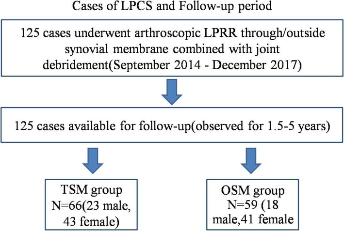
Methods: From September 2014 to December 2017, 125 patients of LPCS underwent arthroscopic LPRR either OSM or TSM combined with joint debridement. In the OSM group, knee joint was cleaned first. The surface of lateral patella retinaculum (LPR) was created the chamber for arthroscopic operation to release LPR. Synovial membrane was retained. In the TSM group, knee joint was cleaned first. Then synovial membrane, joint capsule and LPR, and superficial fascia were gradually incised from the joint cavity to subcutaneous tissue. The synovial membrane was cut open. Before and after surgery, Lysholm score, patella medial shift, Kujala score, VAS score and surgical complications were collected for evaluating clinical overcomes.
Results: All patients were followed up for 1.5-5 years. All patients had significant reduction in knee pain and improved function after 1 month and 1 year. The Lysholm score, the distance of patella medial shift, Kujala score, and VAS score in the OSM group and the TSM group were significantly improved in the final follow-up compared with before surgery (All P < 0.001), but these observed targets before surgery and at the last follow-up were compared between the OSM group and the TSM group with no statistical differences. However, the number of occurrences of joint hematoma and adhesion was significantly higher in the TSM group than the OSM group (P = 0.024).
Conclusions: Arthroscopic closing LPRR for the treatment of LPCS can effectively improve the function and symptoms of patellofemoral joint with the advantages of small trauma, rapid recovery and less complications. But, the number of occurrences of hemarthrosis and joint adhesion were significantly higher in the TSM group than in the OSM group.
Trial registration: The trial registration number (IRCT): IRCT20200205046378N1 and date of registration: February 10, 2020 (retrospectively registered).
Keywords: Arthroscopy; Lateral patella retinaculum releasing; Lateral patellar compression syndrome; Synovial membrane.
Conflict of interest statement
The authors declare that they have no competing interests.
Figures
References
Publication types
MeSH terms
LinkOut - more resources
Full Text Sources
