. 2020 Feb 17;39(1):36.
doi: 10.1186/s13046-020-1542-z.

YAP1 plays a key role of the conversion of normal fibroblasts into cancer-associated fibroblasts that contribute to prostate cancer progression

Affiliations

YAP1 plays a key role of the conversion of normal fibroblasts into cancer-associated fibroblasts that contribute to prostate cancer progression

Tianyu Shen et al. J Exp Clin Cancer Res. .

Abstract

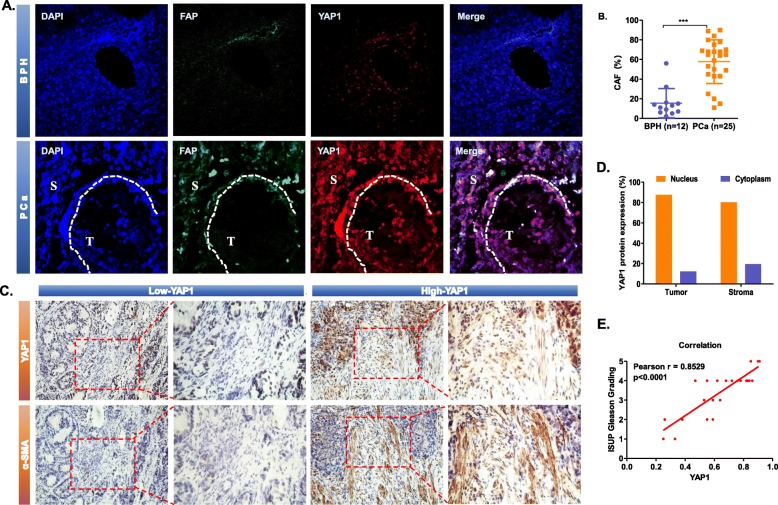
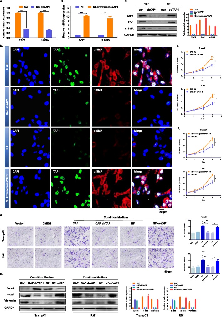
Background: Cancer-associated fibroblasts (CAFs) are an important part of the tumour microenvironment, and their functions are of great concern. This series of experiments aimed to explore how Yes-associated protein 1 (YAP1) regulates the function of stromal cells and how the normal fibroblasts (NFs) convert into CAFs in prostate cancer (PCa).

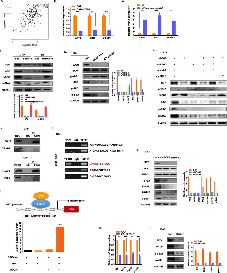
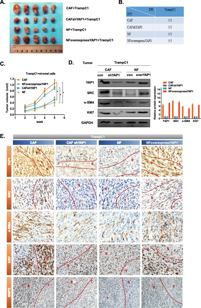
Methods: The effects of conditioned media from different fibroblasts on the proliferation and invasion of epithelial cells TrampC1 were examined. We then analysed the interaction between the YAP1/TEAD1 protein complex and SRC, as well as the regulatory function of the downstream cytoskeletal proteins and actins. A transplanted tumour model was used to explore the function of YAP1 in regulating tumour growth through stromal cells. The relationship between the expression of YAP1 in tumour stromal cells and the clinical characteristics of PCa patients was analysed.

Results: The expression level of YAP1 was significantly upregulated in PCa stromal cells. After the expression level of YAP1 was increased, NF was transformed into CAF, enhancing the proliferation and invasion ability of epithelial cells. The YAP1/TEAD1 protein complex had the capability to influence downstream cytoskeletal proteins by regulating SRC transcription; therefore, it converts NF to CAF, and CAF can significantly promote tumour growth and metastasis. The high expression of YAP1 in the tumour stromal cells suggested a poor tumour stage and prognosis in PCa patients.

Conclusion: YAP1 can convert NFs into CAFs in the tumour microenvironment of PCa, thus promoting the development and metastasis of PCa. Silencing YAP1 in tumour stromal cells can effectively inhibit tumour growth.

Keywords: Cancer-associated fibroblasts (CAFs); Normal fibroblasts (NFs); Prostate cancer (PCa); Yes-associated protein 1 (YAP1).


Conflict of interest statement

The authors declare that they have no competing interests.

Figures

Fig. 1
Fig. 1
Increased expression of YAP1 in stromal cells in PCa. a Immunofluorescence staining showed the protein expression level and location of FAP and YAP1 in BPH (n = 12) and PCa (n = 25). FAP was displayed in red and YAP1 was displayed in green. The nuclei were stained with DAPI and are shown in blue. The representative image had a magnification of 400 x. b Statistical results show that the proportion of CAFs in PCa tissues (n = 25) is significantly higher than that in BPH tissues (n = 12), p < 0.0001. c Immunohistochemical staining showing the expression level and location of YAP1 and α-SMA in PCa (n = 25). Based on the expression of YAP1 in stromal cells, the IHC results were divided into two groups: Low-YAP1 (n = 9) and High-YAP1 (n = 16). The representative image had a magnification of 200 x. d Statistical results showing the localization of YAP1 in the cells of the “YAP1-High” group (n = 16). YAP1 was localized in the nucleus in 87.65% of tumour cells and 80.36% of stromal cells, while it was localized in the cytoplasm in 12.35% of tumour cells and 19.64% of stromal cells. e The correlation between YAP1 expression in prostate cancer stromal cells and the Gleason Grading. The abscissa represents the positive rate of YAP1 in prostate cancer stromal cells, and the ordinate represents the Gleason Grading. Pearson r = 0.8529, p < 0.0001
Fig. 2
Fig. 2
YAP1 plays an important role in the conversion of NFs to CAFs in vitro. a-b The mRNA expression of YAP1 and α-SMA in the CAF, CAFshYAP1, NF and NFoverexpressYAP1 groups were detected by qRT-PCR. c The protein expression of YAP1, FAP and α-SMA in the indicated four cell lines were detected by western blot. GAPDH was used as an endogenous reference gene. d Immunofluorescence staining shows the expression level and location of YAP1 and α-SMA in the four indicated four cells. The nuclei were stained with DAPI. The representative image had a magnification of 400 x. e-f The MTT experiment showing the effect of the conditioned medium on the four indicated cell lines on the proliferation of the epithelial cells TrampC1 or RM1. The absorbance value was detected at a wavelength of 570 nm (*P < 0.05). g The Transwell invasion assay detects the effect of the conditioned medium on the indicated four cell lines on the invasive ability of the epithelial cells TrampC1 or RM1. Statistical results (right side) of the above invasive ability. Five visual field counts were taken for each group, and the ordinate indicates the number of invading cells (***P < 0.001). h The protein expression of E-cad, N-cad and vimentin in the indicated four cell lines were detected by western blot. GAPDH was used as an endogenous reference gene
Fig. 3
Fig. 3
YAP1 activates actin and cytoskeletal proteins to transform NFs to CAFs by regulating SRC. a The association of YAP1 and SRC in prostate cancer was analysed online at http://gepia.cancer-pku.cn/. Pearson R = 0.32. b-c The mRNA expression levels of YAP1, α-SMA and SRC in the indicated four cell lines were detected by qRT-PCR. d The protein expression levels of YAP1, α-SMA, SRC and p-SRC in the indicated four cell lines were detected by western blot. GAPDH was used as an endogenous reference gene. e Western blot was used to detect the protein expression levels of TEAD1, YAP1, p-YAP1, SRC, p-SRC and α-SMA after siTEAD1 transfection of the CAFs. GAPDH was used as an endogenous reference gene. f Western blot was used to detect the protein expression levels of TEAD1, YAP1, p-YAP1, SRC, p-SRC and α-SMA when they were knocked down or overexpressed in CAFs. GAPDH was used as an endogenous reference gene. g The interaction between YAP1 and TEAD1 in the CAFs was determined by the co-IP assay. The relative levels of YAP1 or TEAD1 in these cells were determined by western blot using a YAP1 or TEAD1 antibody. h Chromatin immunoprecipitation (ChIP) of the CAFs was performed with control IgG and TEAD1 antibodies. The precipitation of the SRC promoter was examined by PCR. i A dual-luciferase reporter assay driven by the SRC promoter was co-transfected in the presence or absence of YAP1 or TEAD1. The relative luciferase activities were determined by calculating the ratio of firefly luciferase activities over Renilla luciferase activities. Three independent experiments were conducted, with the means±s.d. of the relative luciferase activities shown. j Western blot was used to detect the protein expression levels of SRC, YAP1, TEAD1, MYL9, F-actin, paxillin and α-SMA after siSRC transfection of the CAFs. GAPDH was used as an endogenous reference gene. k qRT-PCR detection of mRNA expression levels of MYL9, F-actin and paxillin in the CAFshYAP1 group. l Western blot was used to detect the protein expression levels of SRC, MYL9, F-actin and paxillin in the CAFshYAP1 group. GAPDH was used as an endogenous reference gene
Fig. 4
Fig. 4
Fibroblasts with high expression of YAP1 promote tumour growth in vivo. a Tumour photos taken with a digital camera. b Statistical table of the number of tumours in each experimental group. c The line graph shows the subcutaneous tumour volumes as a function of time. The data were recorded from the second week to the fifth week (* P < 0.05). e Western blot was used to detect the expression levels of YAP1, SRC, α-SMA and Ki67 in tumour tissues. GAPDH was used as an endogenous reference gene. f Immunohistochemical staining was used to detect the expression levels and positions of YAP1, SRC, α-SMA, Ki67 and MMP2 in tumour tissues. The tumour cells and stromal cells were separated by curves. T: tumour cells, S: stromal cells
Fig. 5
Fig. 5
Expression of YAP1 in the CAFs of prostate cancer patients. a Immunofluorescence staining for the detection of YAP1 and SRC expression in stromal cells of PCa samples (n = 25). The representative images had a magnification of 400 x. b Correlation between expression of YAP1 and SRC in prostate cancer stromal cells. The abscissa represents the positive rate of YAP1 in prostate cancer stromal cells, and the ordinate represents the positive rate of SRC in prostate cancer stromal cells. Pearson r = 0.7534, P < 0.001. c Primary cell culture was performed by clinical surgical specimens, and the primary hCAF and hNF cells were photographed under a light microscope. The representative images were magnified 200 x. d Immunofluorescence technique was used to detect the expression level and location of α-SMA in hNF and hCAF. The representative images were magnified 400 x. e The interaction between YAP1 and SRC in the hCAF was determined by the co-IP assay. The relative levels of YAP1 or SRC in these cells were determined by western blot using a YAP1 or SRC antibody. f Western blot was used to detect the protein expression levels of YAP1, SRC, FAP and α-SMA in hNF and hCAF. g The protein expression levels of YAP1, SRC and α-SMA were detected after transfection of hCAF with siYAP1. h The protein expression levels of YAP1 and α-SMA were detected after transfection of hCAF with siSRC. GAPDH was used as an endogenous reference gene. i The MTT experiment showing the effect of the conditioned medium on hCAF cell lines on the proliferation of the epithelial cells LNCaP or PC3. The absorbance value was detected at a wavelength of 570 nm (*P < 0.05). j The Transwell invasion assay detects the effect of the conditioned medium on hCAF cell lines on the invasive ability of the epithelial cells LNCaP or PC3. Statistical results (right side) of the above invasive ability. Five visual field counts were taken for each group, and the ordinate indicates the number of invading cells (***P < 0.001)

References

    1. Siegel RL, Miller KD, Jemal A. Cancer statistics, 2018. CA Cancer J Clin. 2018;68(1):7–30. - PubMed
    1. Abylkassov R, Xie Y. Role of yes-associated protein in cancer: an update. Oncol Lett. 2016;12(4):2277–2282. doi: 10.3892/ol.2016.4955. - DOI - PMC - PubMed
    1. Albrengues J, Bertero T, Grasset E, Bonan S, Maiel M, Bourget I, Philippe C, Herraiz Serrano C, Benamar S, Croce O, et al. Epigenetic switch drives the conversion of fibroblasts into proinvasive cancer-associated fibroblasts. Nat Commun. 2015;6:10204. doi: 10.1038/ncomms10204. - DOI - PMC - PubMed
    1. Collak FK, Demir U, Ozkanli S, Kurum E, Zerk PE. Increased expression of YAP1 in prostate cancer correlates with extraprostatic extension. Cancer Biol Med. 2017;14(4):405–413. doi: 10.20892/j.issn.2095-3941.2017.0083. - DOI - PMC - PubMed
    1. Dong A, Gupta A, Pai RK, Tun M, Lowe AW. The human adenocarcinoma-associated gene, AGR2, induces expression of amphiregulin through hippo pathway co-activator YAP1 activation. J Biol Chem. 2011;286(20):18301–18310. doi: 10.1074/jbc.M110.215707. - DOI - PMC - PubMed

MeSH terms