. 2020 Feb 17;10(1):2751.
doi: 10.1038/s41598-020-59539-6.

Characteristics of Atrial Fibrillation Patients Suffering Esophageal Injury Caused by Ablation for Atrial Fibrillation

Affiliations

Characteristics of Atrial Fibrillation Patients Suffering Esophageal Injury Caused by Ablation for Atrial Fibrillation

Pei Zhang et al. Sci Rep. .

Abstract

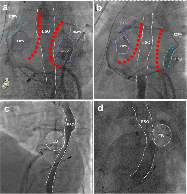
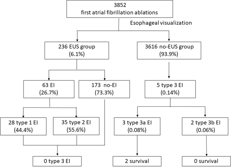
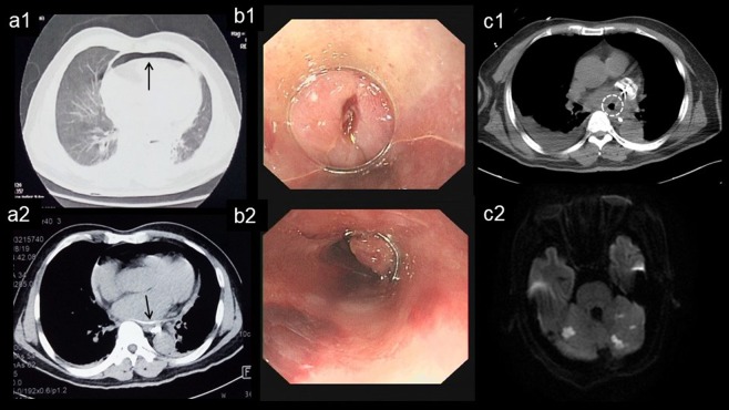
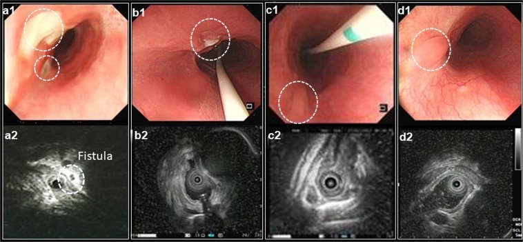
The close proximity of esophagus to the left atrial posterior wall predisposes esophagus to thermal injury during catheter ablation for atrial fibrillation (AF). In this retrospective study, we aimed to investigate risk factors of esophageal injury (EI) caused by catheter ablation for AF. Patients who underwent first-time AF ablation from July 2013 to June 2018 were included. The esophagus was visualized by oral soluble contrast during ablation for all patients and a subset of patients were selected to undergo endoscopic ultrasonography (EUS) to estimate EI post ablation. Degree of EI was categorized as Kansas City classification: type 1: erythema; type 2: ulcers (2a: superficial ulcers; 2b: deep ulcers); type 3: perforation (3a: perforation without communication with the atria; 3b: atrioesophageal fistula [AEF]). Of 3,852 patients, 236 patients (61.5 ± 9.7 years; male, 69%) received EUS (EUS group) and 3616 (63.2 ± 10.9 years; male, 61.1%) without EUS (No-EUS group). In EUS group, EI occurred in 63 patients (type 1 EI in 35 and type 2 EI in 28), and no type 3 EI was observed during follow up. In a multivariable logistic regression analysis, an overlap between the ablation lesion and esophagus was an independent predictor of EI (odds ratio, 21.2; 95% CI: 6.23-72.0; P < 0.001). In No-EUS group, esophagopericardial fistula (EPF; n = 3,0.08%) or AEF (n = 2,0.06%) was diagnosed 4-37 days after ablation. In 3 EPF patients, 2 completely recovered with conservative management and 1 died. Two AEF patients died. Ablation at the vicinity of the esophagus predicts risk of EI. EUS post ablation may prevent the progression of EI and should be considered in management of EI. It remains challenging to identify patients with high risk of EI.


Conflict of interest statement

The authors declare no competing interests.

Figures

Figure 1
Figure 1
Overlap between the ablation lesion and esophagus course. The designed radiofrequency ablation lesion with (a, PA view) and without (b, PA view) overlap with esophagus course. Cryo ablation lesion with (c, LAO view) and without (d, LAO vies) overlap with esophagus course. CB = cryoballoon; ESO = esophagus.
Figure 2
Figure 2
Esophageal injury post ablation shown by endoscopic ultrasonography. Gastroscopy revealed esophageal ulcer (a1, white dot circle) 2 days after radiofrequency ablation. Ultrasonography displayed the thickening and loss of the submucous layers architecture (a2, white dot circle). The esophagus recovered with normal architecture 10 days later (b1, b2, white dot circle).
Figure 3
Figure 3
Flow chart of patients cohort analyzed and included in the study. EI = esophageal injury; EUS = endoscopic ultrasonography; Kansas City classification: type 1: erythema; type 2: ulcers (2a: superficial ulcers; 2b: deep ulcers); type 3: perforation (3a: perforation without communication with the atria; 3b: AEF).
Figure 4
Figure 4
Progression from esophagopericardial fistula to atrioesophageal fistula. Unenhanced CT revealed pneumopericardium (a1, arrow) and the presence of Barium sulfate suspension prior esophagraphy within the pericardial sac (a2, arrow). Endoscopic closure of esophagus perforation with titanium clips (b1,b2). The gas (white dot circle) in left atrium documented by CT (c1) and multiple cerebral embolism documented by MRI (c2).
Figure 5
Figure 5
Evolution of the esophagopericardial fistula post ablation. The gastroscopy demonstrated the progression of esophagopericardial fistula 7 days (a1) 14 days (b1), 21 days (c1) and 30 days (d1) after RF ablation. Ultrasonography revealed dense echo with air, which indicated local fistula formation (a2, white dot circle) and recovery of esophageal tissue layer (b2,c2,d2).

References

    1. Mentias A, Briasoulis A, Shantha G, Alvarez P, Vaughan-Sarrazin M. Impact of Heart Failure Type on Thromboembolic and Bleeding Risk in Patients With Atrial Fibrillation on Oral Anticoagulation. Am J Cardiol. 2019;123:1649–1653. doi: 10.1016/j.amjcard.2019.02.027. - DOI - PMC - PubMed
    1. Cappato R, et al. Prevalence and causes of fatal outcome in catheter ablation of atrial fibrillation. J Am Coll Cardiol. 2009;53:1798–1803. doi: 10.1016/j.jacc.2009.02.022. - DOI - PubMed
    1. Martinek M, et al. Esophageal damage during radiofrequency ablation of atrial fibrillation: impact of energy settings, lesion sets, and esophageal visualization. J Cardiovasc Electrophysiol. 2009;20:726–733. doi: 10.1111/j.1540-8167.2008.01426.x. - DOI - PubMed
    1. Barbhaiya CR, et al. Global survey of esophageal and gastric injury in atrial fibrillation ablation: incidence, time to presentation, and outcomes. J Am Coll Cardiol. 2015;65:1377–1378. doi: 10.1016/j.jacc.2014.12.053. - DOI - PubMed
    1. Halbfass, P. et al. Progression From Esophageal Thermal Asymptomatic Lesion to Perforation Complicating Atrial Fibrillation Ablation: A Single-Center Registry. Circ Arrhythm Electrophysiol10, 10.1161/CIRCEP.117.005233 (2017). - PubMed

MeSH terms