. 2020 Feb 17;10(1):2768.
doi: 10.1038/s41598-020-59639-3.

Muscle Injury Induces Postoperative Cognitive Dysfunction

Affiliations

Muscle Injury Induces Postoperative Cognitive Dysfunction

Lorna Guéniot et al. Sci Rep. .

Abstract

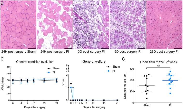
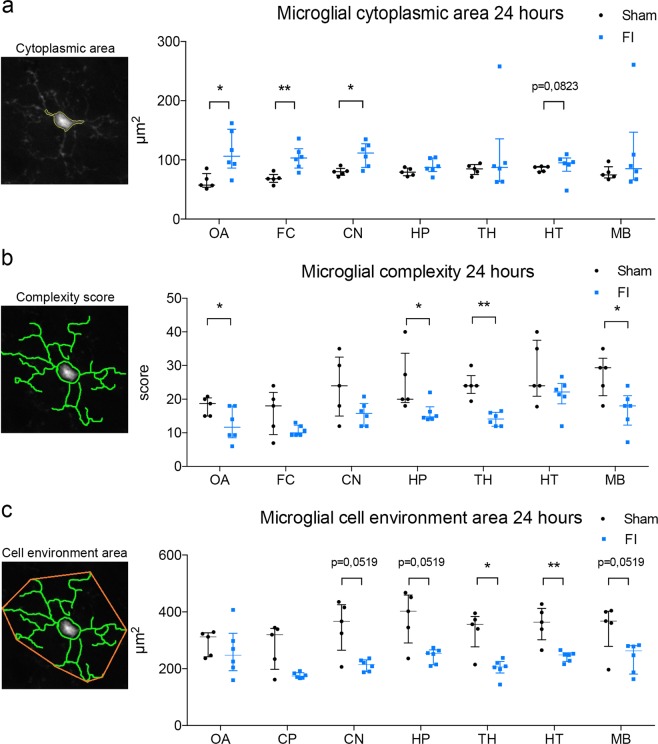
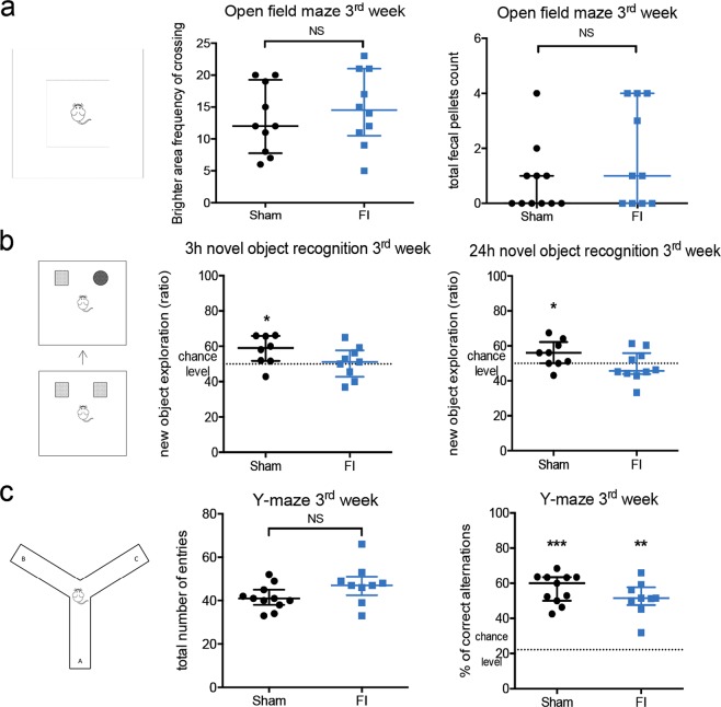
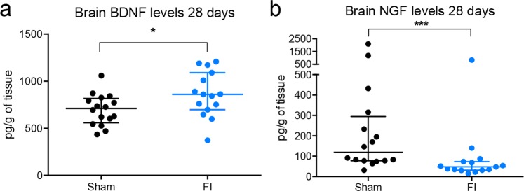
Postoperative cognitive dysfunction (POCD) is a major complication affecting patients of any age undergoing surgery. This syndrome impacts everyday life up to months after hospital discharge, and its pathophysiology still remains unclear. Translational research focusing on POCD is based on a wide variety of rodent models, such as the murine tibial fracture, whose severity can limit mouse locomotion and proper behavioral assessment. Besides, influence of skeletal muscle injury, a lesion encountered in a wide range of surgeries, has not been explored in POCD occurrence. We propose a physical model of muscle injury in CX3CR1GFP/+ mice (displaying green fluorescent microglial cells) to study POCD, with morphological, behavioral and molecular approaches. We highlighted: alteration of short- and long-term memory after muscle regeneration, wide microglial reactivity in the brain, including hippocampus area, 24 hours after muscle injury, and an alteration of central brain derived neurotrophic factor (BDNF) and nerve growth factor (NGF) balance, 28 days after muscle injury. Our results suggest for the first time that muscle injury can have early as well as late impacts on the brain. Our CX3CR1GFP/+ model can also facilitate microglial investigation, more specifically their pivotal role in neuroinflammation and synaptic plasticity, in the pathophysiology of POCD.


Conflict of interest statement

The authors declare no competing interests.

Figures

Figure 1
Figure 1
Muscle freeze-injury induces only mild general disturbance and muscle regeneration within 3 weeks in young adult male CX3CR1GFP/+ mice. (a) Hematoxylin-Eosin staining on cryosections. Sham mice did not display any skeletal muscle lesion in these procedures. In contrast, muscle freeze injury (FI) induced necrosis of muscle tissue (24 h post-injury), and regeneration (completed 28 days post-injury, with the remaining of central nuclei). Scale bar = 100 µm. (b) General impact of FI was characterized by weight preservation within 3 weeks after the procedure, and by moderate lameness observed for all FI mice in the first 24 hours among criteria of welfare score (Table 1) resulting in a score at 1/8. All Sham mice get the best welfare score (0/8), during the entire protocol period (n = 11 for Sham group, n = 10 for FI group). (c) Muscle functional recovery was characterized by similar locomotion between FI and Sham group in open field evaluation, 21 days after the surgery (n = 10 for each group). Data are represented as median with inter-quartile range. Significance is indicated with asterisk (**p < 0.01, NS: non-significant); analyzed with Mann-Whitney U test.
Figure 2
Figure 2
Freeze-injury induced widespread microglial morphological changes in brain 24 hours after surgery in young adult male CX3CR1GFP/+ mice. Automated morphometric analysis of microglial cells in olfactive area (OA), frontal cortex (FC), central nuclei (CN), hippocampus (HP), thalamus (TH), hypothalamus (HP) and midbrain (MB). (a) Microglial cytoplasmic area was significantly increased in OA, FC and CN for FI group compared to Sham. The same tendency was observed in HT. (b) Microglial complexity score, reflecting the ramification of microglial cells, was significantly decreased in OA, HP, TH and MB for FI group compared to Sham. (c) Microglial cell environment area was significantly decreased in TH and HT for FI group compared to Sham. The same tendency was observed in CN, HP and MB. (n = 5 for Sham group, n = 6 for FI group). Data are represented as median with inter-quartile range. Significance is indicated with asterisk (*p < 0.05, **p < 0.01); analyzed with Mann-Whitney U test.
Figure 3
Figure 3
Freeze-injury induced cognitive impairments 3 weeks after surgery in young adult CX3CR1GFP/+ mice. (a) Before evaluating memory process in both groups, level of anxiety was controlled during open field maze test to avoid any interference in the following memory evaluations. No differential anxiety was revealed between Sham and FI groups (n = 10 for Sham group in crossing frequency of brighter area and 11 in total fecal pellet count, n = 10 for FI group). (b) Novel object recognition (NOR) test was used to assess either short- and long-term memory. Success in this test is defined by a group score above the level chance (50%). FI group failed in the memory evaluation either at 3 hours (med 51% [IQR 43–58] p = 0.8070) or 24 hours (med 46% [IQR 41–60] p = 0.9821), in contrast to Sham group (3 h NOR: med 59% [IQR 52–66] p = 0.0391, n = 8 for Sham group and n = 9 for FI group; 24 h NOR: med 56% [IQR 49–61] p = 0.0195, n = 9 for Sham group and n = 10 for FI group). (c) To investigate another type of memorization process, immediate spatial memory was evaluated using spontaneous alternation test (Y-maze test). Total number of entries in all arms was similar in Sham and FI mice. Both groups succeeded as they were significantly above the chance level of 20% (n = 11 for Sham group, n = 9 for FI group). Data are represented as median with inter-quartile range. Significance is indicated with asterisk (*p < 0.05, **p < 0.01, ***p < 0.001); analyzed with Wilcoxon Signed Rank test for median comparison with 50% and 22.2%, analyzed with Mann-Whitney U test for group comparison. Mouse drawing by Clker-Free-Vector-Images from Pixabay.
Figure 4
Figure 4
Freeze-injury altered brain levels of neurotrophins 3 weeks after surgery in young adult CX3CR1GFP/+ mice. On the same animal cohort presenting differential cognitive performances, levels of brain derived neurotrophic factor (BDNF) and nerve growth factor (NGF) were measured in brain homogenate 28 days after surgical procedure. (a) Brain levels of BDNF were significantly increased in FI mice (med 860 pg/g of tissue [IQR 700–1090]) compared to Sham (med 710 pg/g of tissue [IQR 560–820]; p = 0.0296) in ELISA assay. (b) Brain levels of NGF were significantly decreased in FI mice (med 48 pg/mL [IQR 31–74]) compared to Sham (med 119 pg/mL [77–295]; p = 0.0009) in multiplex assay. Data are represented as median with inter-quartile range (n = 15 for each group). Significance is indicated with asterisk (*p < 0.05, ***p < 0.001); analyzed with Mann-Whitney U test.

References

    1. Monk TG, et al. Predictors of cognitive dysfunction after major noncardiac surgery. Anesthesiology. 2008;108:18–30. doi: 10.1097/01.anes.0000296071.19434.1e. - DOI - PubMed
    1. Krenk L, Rasmussen LS, Kehlet H. New insights into the pathophysiology of postoperative cognitive dysfunction. Acta Anaesthesiol. Scand. 2010;54:951–6. doi: 10.1111/j.1399-6576.2010.02268.x. - DOI - PubMed
    1. Czyż-Szypenbejl K, Mędrzycka-Dąbrowska W, Kwiecień-Jaguś K, Lewandowska K. The Occurrence of Postoperative Cognitive Dysfunction (POCD) - Systematic Review. Psychiatr. Pol. 2019;53:145–160. doi: 10.12740/PP/90648. - DOI - PubMed
    1. Evered LA, Silbert BS. Postoperative Cognitive Dysfunction and Noncardiac Surgery. Anesth. Analg. 2018;127:496–505. doi: 10.1213/ANE.0000000000003514. - DOI - PubMed
    1. Rundshagen I. Postoperative cognitive dysfunction. Dtsch. Arztebl. Int. 2014;111:119–25. - PMC - PubMed

Publication types

MeSH terms