Escherichia coli-based synthesis of cadmium sulfide nanoparticles, characterization, antimicrobial and cytotoxicity studies
- PMID: 32067210
- PMCID: PMC7455616
- DOI: 10.1007/s42770-020-00238-9
Escherichia coli-based synthesis of cadmium sulfide nanoparticles, characterization, antimicrobial and cytotoxicity studies
Abstract
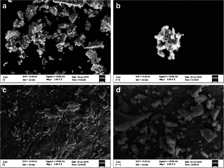
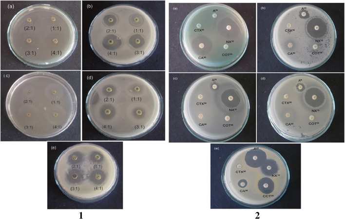
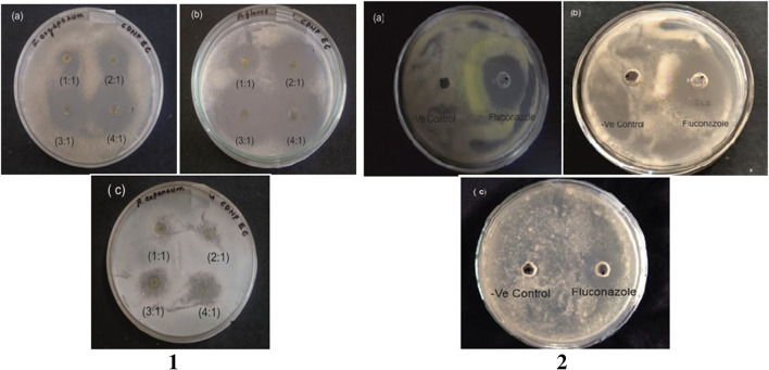
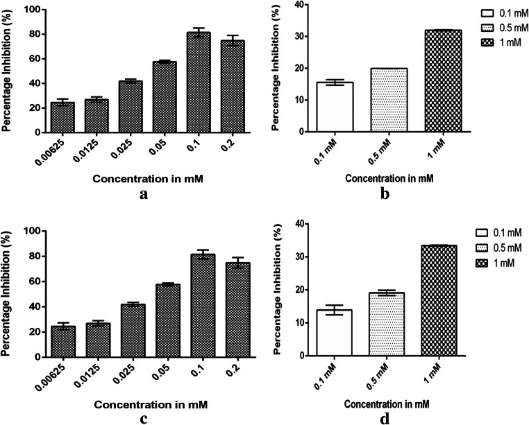
The present research describes the synthesis of cadmium sulfide (CdS) nanoparticles from Escherichia coli under the influence of bacterial enzyme sulphate reductase and study on their cytotoxicity for applications in cancer therapy. Escherichia coli cells were used to synthesize CdS nanoparticles under different concentrations of cadmium chloride and sodium sulfide. The morphology of the nanoparticles was analysed using scanning electron microscopy (SEM) and energy dispersive X-ray spectroscopy (EDX) was used for elemental analysis of nanoparticles. Fourier-transform infrared spectroscopy analysis (FTIR) was performed to assess the functional groups of the nanoparticles. Crystalline nature of nanoparticles was assessed using powder X-ray diffraction (XRD). Antibacterial studies of CdS nanoparticles were carried out on foodborne pathogens and cytotoxicity studies were carried out on Mus musculus skin melanoma (B16F10) and human epidermoid carcinoma (A431) cell lines. CdS nanoparticle showed more cytotoxic effect on cancer cells compared with standard 5-aminolevulinic acid (5-ALA). The Escherichia coli-synthesized CdS nanoparticles showed highest zone of inhibition in the ratio 4:1 of cadmium chloride and sodium sulfide on all tested bacterial strains. The nanoparticles were also tested for haemolytic activity on RBC cells, which exhibited lower cytotoxicity than sodium dodecyl sulphate which was used as positive control. The cytotoxicity of CdS nanoparticles assessed on A431 cells showed an inhibition of 81.53% at 100 μM concentration while the cytotoxicity assessed on B16F10 cells showed an inhibition of 75.71% at 200 μM concentration which was much efficient than 5-ALA which showed an inhibition of 31.95% at a concentration against B16F10 cells and 33.45% against A431 cells at a concentration of 1 mM. Cadmium sulfide nanoparticles were thus found to be highly toxic on cancer cells compare with standard anticancerous drug 5-ALA.
Keywords: 5-Aminolevulinic acid; Antimicrobial activity; Cadmium sulfide nanoparticles; Cytotoxicity.
Conflict of interest statement
The authors declare that they have no conflict of interest.
Figures

References
-
- Mohanapuria P, Rana NK, Yadav SK. Biosynthesis of nanoparticles: technological concepts and future application. J Nanoparticles Res. 2008;10:507–517.
-
- Debaditya B, Rajinder K. Nanotechnology and potential for microorganisms. Crit Rev Biotechnol. 2005;25(4):199–204. - PubMed
-
- Kalishwaralal K, Babu RS, Venkataraman D, Mohd B, Gurunathan S. Biosynthesis of silver nanocrystals by Bacillus licheniformis. Colloids Surf B: Biointerfaces. 2008;65(1):150–153. - PubMed
-
- Singh BR, Dwivedi S, Al-Khedhairy AA, Musarrat J. Colloids and surfaces. B Biointerfaces. 2011;85(2):207–213. - PubMed
MeSH terms
Substances
LinkOut - more resources
Full Text Sources