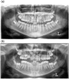
Prediction of maxillary canine impaction based on panoramic radiographs
- PMID: 32067406
- PMCID: PMC7025989
- DOI: 10.1002/cre2.246
Prediction of maxillary canine impaction based on panoramic radiographs
Abstract
Objectives: The objective of this article is to establish a large sample-based prediction model for maxillary canine impaction based on linear and angular measurements on panoramic radiographs and to validate this model.
Materials and methods: All patients with at least two panoramic radiographs taken between the ages of 7 and 14 years with an interval of minimum 1 year and maximum 3 years (T1 and T2) were selected from the Department of Oral Health Sciences, University Hospital Leuven database. Linear and angular measurements were performed at T1. From 2361 records, 572 patients with unilateral or bilateral canine impaction were selected at T1. Of those, 306 patients were still untreated at T2 and were used as study sample. To construct the prediction model, logistic regression analysis was used.
Results: The parameters analyzed through backward selection procedure were canine to midline angle, canine to first premolar angle, canine cusp to midline distance, canine cusp to maxillary plane distance, sector, quadratic trends for continuous predictors, and all pairwise interactions. The final model was applied to calculate the likelihood of impaction and yielded an area under the curve equal to 0.783 (95% CI [0.742-0.823]). The cut-off point was fixed on 0.342 with a sensitivity of 0.800 and a specificity of 0.598. The cross-validated area under the curve was equal to 0.750 (95% CI [0.700, 0.799]).
Conclusion: The prediction model based on the above mentioned parameters measured on panoramic radiographs is a valuable tool to decide between early intervention and regular follow-up of impacted canines.
Keywords: cuspid; impacted tooth; panoramic radiography; predictor.
© 2019 The Authors. Clinical and Experimental Dental Research published by John Wiley & Sons Ltd.
Conflict of interest statement
The authors certify no potential conflicts of interest with respect to the authorship and/or publication of this article.
Figures


References
-
- Alqerban, A. , Jacobs, R. , Fieuws, S. , Nackaerts, O. , & Willems, G. (2011). Comparison of 6 cone‐beam computed tomography systems for image quality and detection of simulated canine impaction‐induced external root resorption in maxillary lateral incisors. Am J Orthod Dentofac Orthop., 140(3), 129–139. - PubMed
Publication types
MeSH terms
LinkOut - more resources
Full Text Sources

