. 2020 May;15(5):777-791.
doi: 10.1016/j.jtho.2020.01.009. Epub 2020 Feb 15.

STING Pathway Expression Identifies NSCLC With an Immune-Responsive Phenotype

Affiliations

STING Pathway Expression Identifies NSCLC With an Immune-Responsive Phenotype

Carminia M Della Corte et al. J Thorac Oncol. 2020 May.

Abstract

Introduction: Although the combination of anti-programmed cell death-1 or anti-programmed cell death ligand-1 (PD-L1) with platinum chemotherapy is a standard of care for NSCLC, clinical responses vary. Even though predictive biomarkers (which include PD-L1 expression, tumor mutational burden, and inflamed immune microenvironment) are validated for immunotherapy, their relevance to chemoimmunotherapy combinations is less clear. We have recently reported that activation of the stimulator of interferon genes (STING) innate immune pathway enhances immunotherapy response in SCLC. Here, we hypothesize that STING pathway activation may predict and underlie predictive correlates of antitumor immunity in NSCLC.

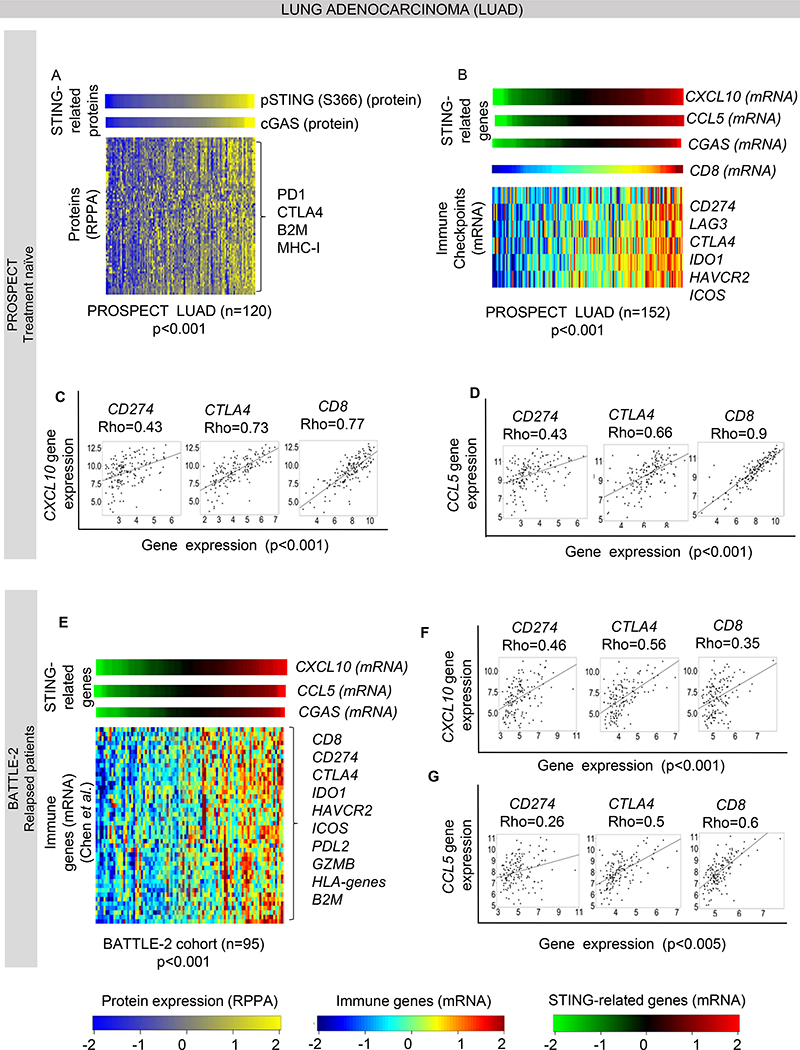
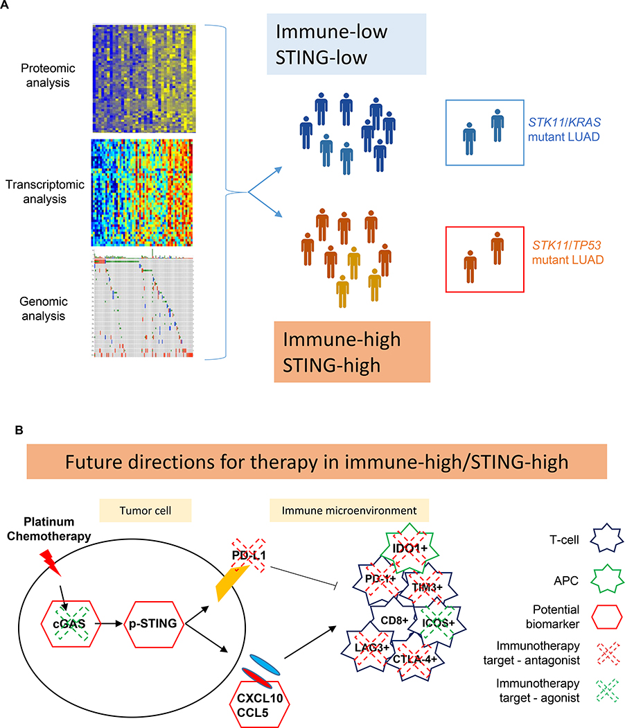
Methods: We analyzed transcriptomic and proteomic profiles in two NSCLC cohorts from our institution (treatment-naive patients in the Profiling of Resistance Patterns and Oncogenic Signaling Pathways in Evaluation of Cancers of the Thorax study and relapsed patients in the Biomarker-Integrated Approaches of Targeted Therapy for Lung Cancer Elimination study) and The Cancer Genome Atlas (N = 1320). Tumors were stratified by STING activation on the basis of protein or mRNA expression of cyclic GMP-AMP synthase, phospho-STING, and STING-mediated chemokines (chemokine ligand 5 [CCL5] and C-X-C motif chemokine 10 [CXCL10]). STING activation in patient tumors and in platinum-treated preclinical NSCLC models was correlated with biomarkers of immunotherapy response.

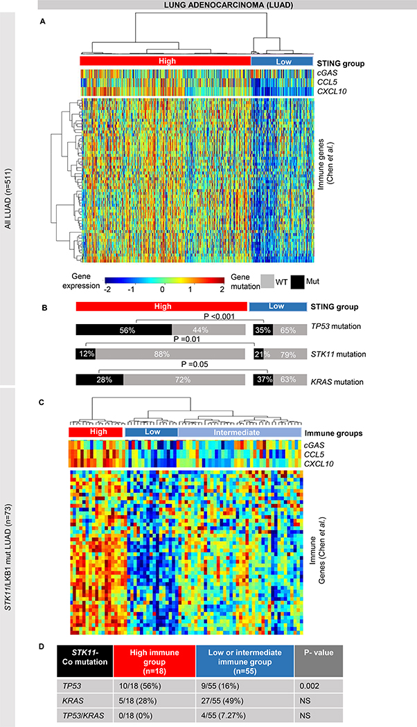
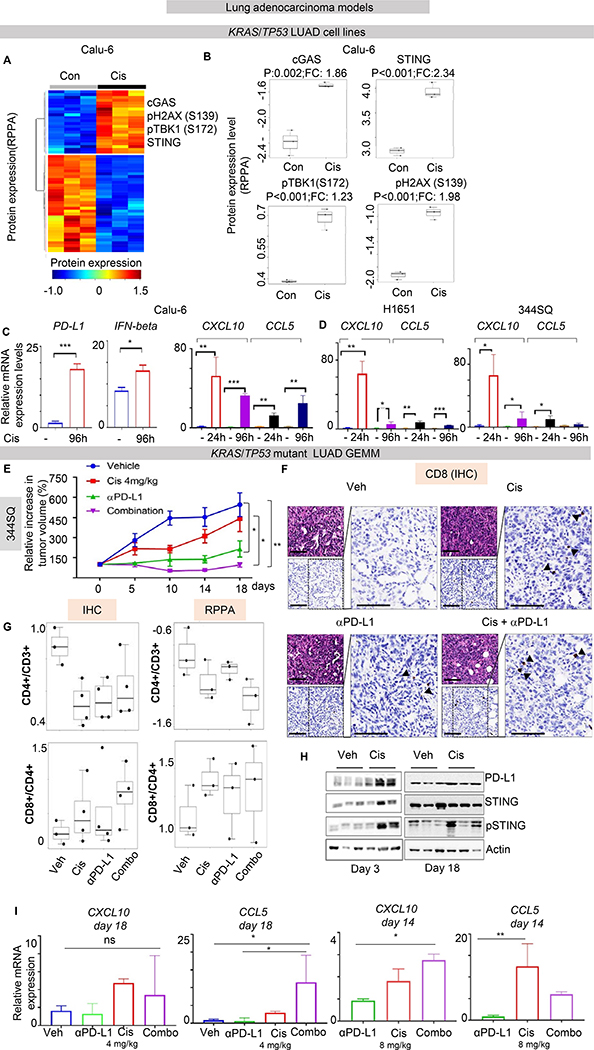
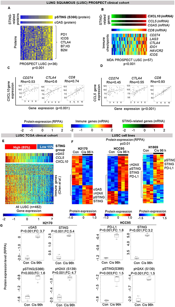
Results: STING activation is associated with higher levels of intrinsic DNA damage, targetable immune checkpoints, and chemokines in treatment-naive and relapsed lung adenocarcinoma. We observed that tumors with lower STING and immune gene expression show higher frequency of serine-threonine kinase 11 (STK11) mutations; however, we identified a subset of these tumors that are TP53 comutated and display high immune- and STING-related gene expression. Treatment with cisplatin increases STING pathway activation and PD-L1 expression in multiple NSCLC preclinical models, including adeno- and squamous cell carcinoma.

Conclusions: STING pathway activation in NSCLC predicts features of immunotherapy response and is enhanced by cisplatin treatment. This suggests a possible predictive biomarker and mechanism for improved response to chemoimmunotherapy combinations.

Keywords: Immune checkpoints; Immunotherapy; Innate immunity; Lung cancer; STING.


Figures

Figure 1.
Figure 1.. STING pathway proteins expression is associated with immune activation in lung adenocarcinoma.
(A) Heatmap shows correlation of phospho-STING (Ser_366) protein levels with CGAS and other immune related proteins in the MD Anderson PROSPECT cohort, including treatment naïve adenocarcinoma (n=120, Spearman’s rho > 0.3, p<0.001). (B) Expression of the STING downstream chemokine CXCL10 is correlated with the other STING related genes CCL5 and CGAS (p<0.001), with expression of selected targetable immune genes: CD274 (PD-L1), LAG3, CTLA4, IDO1, HAVCR2 and ICOS (Spearman’s rho > 0.5, p<0.001) and with CD8 mRNA levels, indicative of immune infiltration in PROSPECT LUAD cohort (n=120). (C-D) Individual dot-plots showing correlation of CXCL10 (C) and CCL5 (D) gene expression with selected targetable genes CD274 and CTLA4, and CD8 in PROSPECT LUAD cohort (n=120). (E) Correlation of CXCL10 with CCL5 and CGAS gene expression, with selected targetable immune genes and other immune markers from the list of Chen et al. in the treatment-refractory and relapsed BATTLE-2 cohort (n=95, Spearman’s rho > 0.3, p<0.001). (F-G) Individual dot-plots showing correlation of CXCL10 (F) and CCL5 (G) gene expression with selected targetable genes CD274 and CTLA4, and CD8 in BATTLE-2 cohort.
Figure 2.
Figure 2.. STK11/LKB1 mutation is associated with different phenotypes of immune activation.
(A-B) Identification of STING/immune signatures genes (from Chen et al.) shows two main groups of LUAD carcinomas in TCGA LUAD cohort (n=515), a low STING/low immune group and a high STING/high immune group (A). We found an enrichment of STK11 (LKB1) (p=0.01) and of KRAS (p=0.05) mutations in low STING/low immune group and of TP53 mutations (p<0.001) in high STING/high immune tumors (B). (C-D) Hierarchical clustering of immune signatures genes (from Chen et al.) in STK11- mutant tumors in the TCGA LUAD cohort identifies three subgroups with low, intermediate and high expression of immune and STING genes (C). Comparison of the three subgroups detected a significant enrichment in the frequency of TP53 mutations (p=0.002, by Fisher’s exact test) in the “immune-high” tumors as compared to the other two subgroups (D). NS= not significant.
Figure 3.
Figure 3.. Cisplatin treatment activates STING pathway markers in NSCLC in vitro and in vivo.
(A-B) Supervised hierarchical clustering of protein expression profiles in untreated and cisplatin-treated LUAD cells Calu-6 (KRAS/TP53 mutant) shows upregulation of proteins in the STING pathway (STING, phospho-TBK1 (Ser_172), CGAS), PD-L1 and phospho-H2AX(Ser_139) following cisplatin treatment (p<0.01, by t-test). (C-D) Quantitative PCR (qPCR) measurement of PD-L1, CCL5, CXCL10 and IFNβ mRNA expression in three KRAS/TP53 mutant NSCLC cell lines treated with cisplatin for 24 and 96 hours. Data presented as mean ±SD and p values by t-test ***p<0.001, **p<0.01, *p<0.05.. (E) 344SQ tumor bearing mice were treated with vehicle, cisplatin (4 mg/kg, i.p., 1/7), anti-PD-L1 antibody (300μg, i.p., 1/7), or the combination. Relative tumor volumes (mean ± S.E.M.) are shown. The combination of cisplatin and anti-PD-L1 antibody significantly potentiated the anti-tumor response to either single agent (P values were calculated by ANOVA, followed by Tukey’s test. (F) Histological analysis of 344SQ tumors demonstrate the presence of CD8+ immune cells. Scale bar = 200 μM. (G) Results from IHC and RPPA analysis for T-cells infiltration: CD8+/CD4+ and CD4+/CD3+ ratio are presented. Ratio were calculated from number of positive cells for any marker/ mm2. CD4+/CD3+ ratio was decreased by IHC (p=0.038 calculated by ANOVA, followed by Dunnett’s test versus vehicle, p=0.02 for cisplatin, p=0.03 for anti-PDL1, p=0.098 for combination) and by RPPA (p=0.1 by ANOVA). Conversely, CD8+/CD4+ ratio was increased by IHC in 2/4 tumors from cisplatin and 3/4 tumors from combination arm (p>0.1 by ANOVA) and the same result were confirmed by RPPA (p>0.1 by ANOVA). (H) Images from western blot analysis on lysates from tumors harvested from the 344SQ tumor bearing mice after three days or at the end of treatment showed an increase in PD-L1 and phospho-STING levels in cisplatin-treated tumors compared to the vehicle group. (I) Quantitative PCR (qPCR) measurement of CCL5 and CXCL10 mRNA expression in tumors after indicated treatments. Data presented as mean ±SD and p values were calculated by ANOVA, followed by Tukey’s test. ***p<0.001, **p<0.01, *p<0.05. ns=not significant.
Figure 4.
Figure 4.. STING pathway activation is associated with expression of immune markers in squamous cell lung carcinoma.
(A) Heatmap shows correlation of phospho-STING with cGAS protein levels and other immune and inflammatory proteins in the MDACC PROSPECT lung squamous cohort (Spearman’s rho > 0.5, p<0.001). (B) Expression of CXCL10 is correlated with other STING genes, CCL5 and CGAS, CD8, and selected targetable immune genes in PROSPECT LUSC: CD274 (PD-L1) (Spearman’s rho = 0.4, p<0.001), LAG3, CTLA4, IDO1, HAVCR2 and ICOS (Spearman’s rho > 0.6, p<0.0001; respectively). (C-D) Individual dot-plots showing correlation of CXCL10 and CCL5 expression with selected targetable genes shown in 4B. (E) Hierchical clustering, as shown for LUAD in Figure 2A, was applied to TCGA LUSC cohort. (F-G) Supervised hierarchical clustering of protein expression profiles in three untreated and cisplatin-treated (3μM for 96hours) LUSC cells shows upregulation of proteins in the STING pathway, PD-L1 and γH2AX following cisplatin treatment (p<0.01, by t-test).
Figure 5.
Figure 5.. Working model.
(A) Schematic representation of the resources used in this work: genomic, transcriptomic and proteomic analysis led to identification of two main group of STING/immune expression in NSCLC, with distinct molecular subgroups in LUAD. (B) Schematic representation of the novel potential therapeutic implication for high STING/high immune NSCLC.

Comment in

References

    1. Ettinger DS, Aisner DL, Wood DE, Akerley W, Bauman J, Chang JY, et al. NCCN Guidelines Insights: Non-Small Cell Lung Cancer, Version 5.2018. J Natl Compr Canc Netw 2018;16:807–21. - PubMed
    1. Hellmann MD, Ciuleanu TE, Pluzanski A, Lee JS, Otterson GA, Audigier-Valette C, et al. Nivolumab plus Ipilimumab in Lung Cancer with a High Tumor Mutational Burden. N Engl J Med 2018;378:2093–104. - PMC - PubMed
    1. Reck M, Rodríguez-Abreu D, Robinson AG, Hui R, Csőszi T, Fülöp A, et al. Pembrolizumab versus Chemotherapy for PD-L1-Positive Non-Small-Cell Lung Cancer. N Engl J Med 2016;375:1823–33. - PubMed
    1. Skoulidis F, Goldberg ME, Greenawalt DM, Hellmann MD, Awad MM, Gainor JF, et al. STK11/LKB1 Mutations and PD1 Inhibitor Resistance in KRAS-Mutant Lung Adenocarcinoma. Cancer Discov 2018; 8:822–35. - PMC - PubMed
    1. Lee CK, Man J, Lord S, Links M, Gebski V, Mok T, et al. Checkpoint Inhibitors in Metastatic EGFR-Mutated Non-Small Cell Lung Cancer-A Meta-Analysis. J Thorac Oncol 2017;12:403–7. - PubMed

Publication types