

Characterization of the inflammatory features of central centrifugal cicatricial alopecia
- PMID: 32068905
- PMCID: PMC7401378
- DOI: 10.1111/cup.13666
Characterization of the inflammatory features of central centrifugal cicatricial alopecia
Abstract
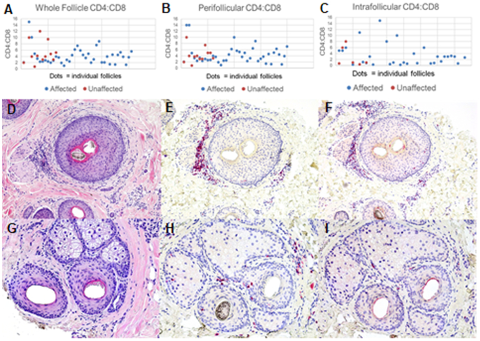
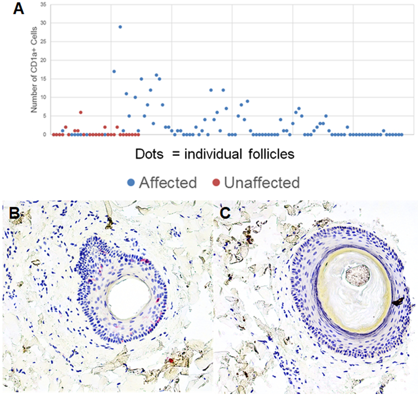
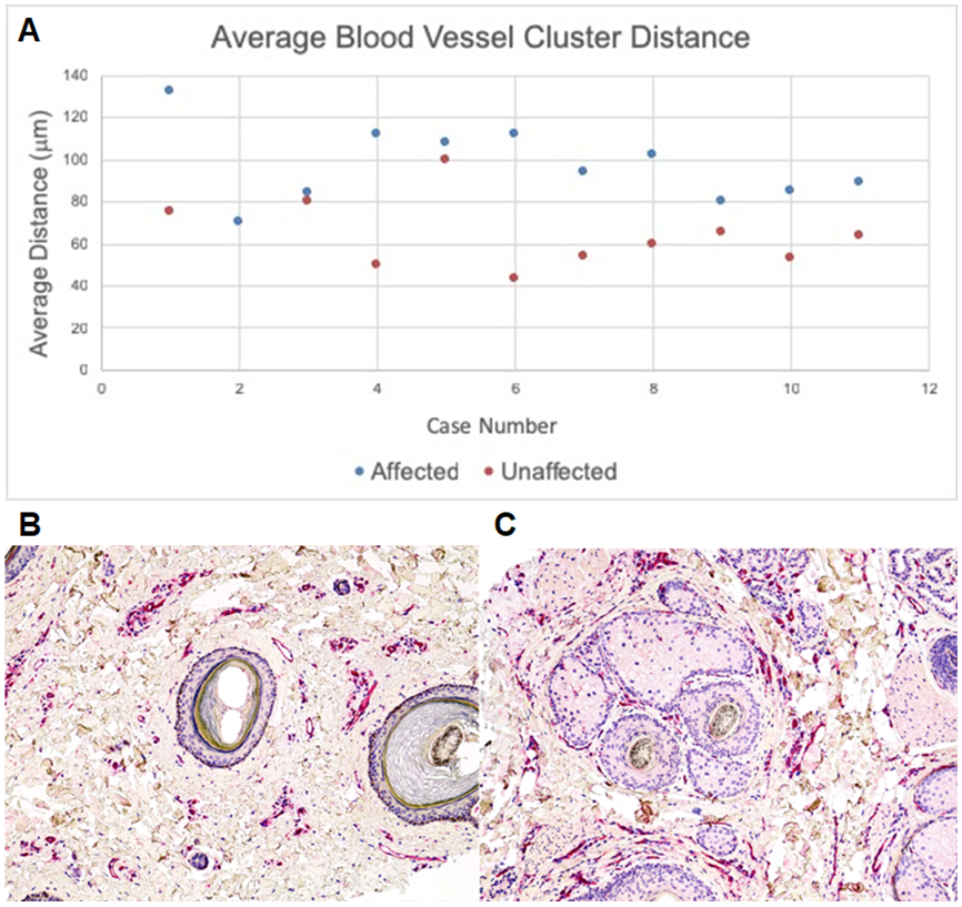
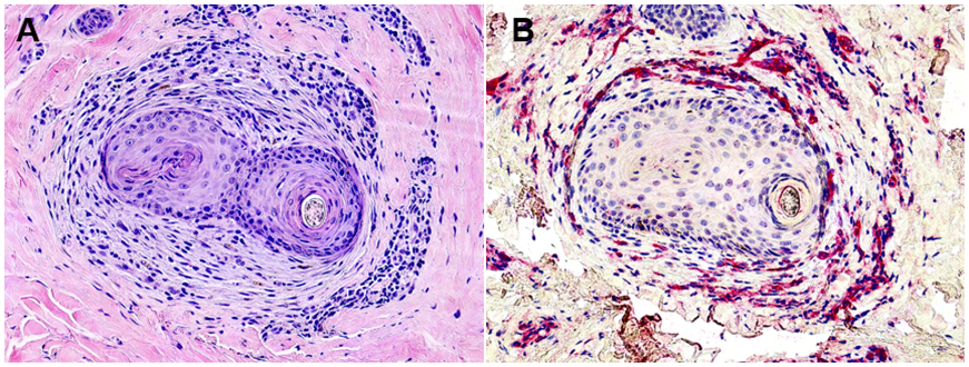
Central centrifugal cicatricial alopecia (CCCA) is a scarring alopecia that primarily affects women of African descent. Although histopathological features of CCCA have been described, the pathophysiology of this disease remains unclear. To better understand the components of CCCA pathophysiology, we evaluated the composition of the inflammatory infiltrate, the distribution of Langerhans cells (LCs), and the relationship between fibrosis and perifollicular vessel distribution. Our data indicate that CCCA is associated with a CD4-predominant T-cell infiltrate with increased LCs extending into the lower hair follicle. Fibroplasia associated with follicular scarring displaces blood vessels away from the outer root sheath epithelium. These data indicate that CCCA is an inflammatory scarring alopecia with unique pathophysiologic features that differentiate it from other lymphocytic scarring processes.
Keywords: CD4 T-cells; Langerhans cells; central centrifugal; cicatricial alopecia; scarring alopecia.
© 2020 John Wiley & Sons A/S. Published by John Wiley & Sons Ltd.
Figures
Similar articles
-
Inflammatory features of frontal fibrosing alopecia.J Cutan Pathol. 2017 Aug;44(8):672-676. doi: 10.1111/cup.12955. Epub 2017 Jun 1. J Cutan Pathol. 2017. PMID: 28429464 Free PMC article. Clinical Trial.
-
Central Centrifugal Cicatricial Alopecia: Challenges and Treatments.Dermatol Clin. 2021 Jul;39(3):389-405. doi: 10.1016/j.det.2021.03.004. Epub 2021 May 15. Dermatol Clin. 2021. PMID: 34053593 Review.
-
Pathologic diagnosis of central centrifugal cicatricial alopecia on horizontal sections.Am J Dermatopathol. 2014 Nov;36(11):859-64; quiz 865-7. doi: 10.1097/DAD.0000000000000174. Am J Dermatopathol. 2014. PMID: 25222198
-
Investigation of the hair follicle inner root sheath in scarring and non-scarring alopecia.J Cutan Pathol. 2007 Oct;34(10):762-8. doi: 10.1111/j.1600-0560.2006.00696.x. J Cutan Pathol. 2007. PMID: 17880581
-
A proposed mechanism for central centrifugal cicatricial alopecia.Exp Dermatol. 2020 Feb;29(2):190-195. doi: 10.1111/exd.13664. Epub 2018 Jun 29. Exp Dermatol. 2020. PMID: 29660185 Review.
Cited by
-
Scalp microbiome: a guide to better understanding scalp diseases and treatments.Arch Dermatol Res. 2024 Jul 29;316(8):495. doi: 10.1007/s00403-024-03235-2. Arch Dermatol Res. 2024. PMID: 39073596 Review.
-
Disruption of the endopeptidase ADAM10-Notch signaling axis leads to skin dysbiosis and innate lymphoid cell-mediated hair follicle destruction.Immunity. 2021 Oct 12;54(10):2321-2337.e10. doi: 10.1016/j.immuni.2021.09.001. Epub 2021 Sep 27. Immunity. 2021. PMID: 34582748 Free PMC article.
-
Dermal T cell immunity and key regulatory signaling pathways: Implications in immune-mediated alopecia and hair regeneration.Genes Dis. 2025 Jan 8;12(5):101518. doi: 10.1016/j.gendis.2025.101518. eCollection 2025 Sep. Genes Dis. 2025. PMID: 40520999 Free PMC article. Review.
-
Topical Treatment for Scarring and Non-Scarring Alopecia: An Overview of the Current Evidence.Clin Cosmet Investig Dermatol. 2021 May 12;14:485-499. doi: 10.2147/CCID.S284435. eCollection 2021. Clin Cosmet Investig Dermatol. 2021. PMID: 34012282 Free PMC article. Review.
-
Immune-mediated alopecias and their mechanobiological aspects.Cells Dev. 2022 Jun;170:203793. doi: 10.1016/j.cdev.2022.203793. Epub 2022 May 29. Cells Dev. 2022. PMID: 35649504 Free PMC article. Review.
References
-
- Olsen EA, Callender V, McMichael A, et al. Central hair loss in African American women: incidence and potential risk factors. Journal of the American Academy of Dermatology. 2011;64(2):245–252. - PubMed
-
- Callender VD, Wright DR, Davis EC, Sperling LC. Hair breakage as a presenting sign of early or occult central centrifugal cicatricial alopecia: clinicopathologic findings in 9 patients. Archives of Dermatology. 2012;148(9):1047–1052. - PubMed
-
- Miteva M. A comprehensive approach to hair pathology of horizontal sections. The American Journal of Dermatopathology. 2013;35(5):529–540. - PubMed
-
- Miteva M, Torres F, Tosti A. The 'eyes' or 'goggles' as a clue to the histopathological diagnosis of primary lymphocytic cicatricial alopecia. The British Journal of Dermatology. 2012;166(2):454–455. - PubMed
-
- Miteva M, Tosti A. 'A detective look' at hair biopsies from African-American patients. The British Journal of Dermatology. 2012;166(6):1289–1294. - PubMed
MeSH terms
Grants and funding
LinkOut - more resources
Full Text Sources
Medical
Research Materials
