. 2020 Mar;294(3):714-715.
doi: 10.1148/radiol.2019181548.

Case 279

Affiliations

Case 279

Peter Abraham et al. Radiology. 2020 Mar.

Abstract

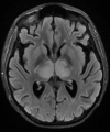
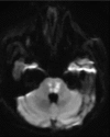
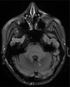
HistoryA 25-year-old woman with recently diagnosed systemic lupus erythematosus and class IV lupus nephritis confirmed with biopsy and treated with mycophenolate mofetil presented with a 2-day history of progressively worsening edema of her face and lower extremities. She had no antecedent infection or vaccination. She was admitted to the hospital and treated with methylprednisolone, furosemide, and C1 esterase inhibitor. On hospital day 2, she experienced a witnessed generalized tonic-clonic seizure. At that time, she became hypoxic and was intubated for airway protection. Her laboratory study results preceding the seizure were remarkable for hyponatremia, with a blood sodium level of 122 mEq/L (122 mmol/L) (normal range, 135-145 mEq/L [134-145 mmol/L]), which was corrected to 137 mEq/L (137 mmol/L) over 48 hours. Same-day cerebrospinal fluid analysis was unremarkable, and unenhanced head CT findings (not shown) were normal, with no evidence of intracranial hemorrhage or edema.


Figures
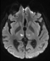
Figure 1a:
Figure 1a:
(a, d) Axial diffusion-weighted MRI (repetition time msec/echo time msec, 6887/76; b value = 1000 msec). (b, e) Axial T2-weighted fluid-attenuated inversion recovery MRI (repetition time msec/echo time msec/inversion time msec, 11 000/99/2650). (c, f) Axial susceptibility-weighted MRI (51/24). All images were obtained at the level of the thalami (a, b, c) and pons (d, e, f).
Figure 1b:
Figure 1b:
(a, d) Axial diffusion-weighted MRI (repetition time msec/echo time msec, 6887/76; b value = 1000 msec). (b, e) Axial T2-weighted fluid-attenuated inversion recovery MRI (repetition time msec/echo time msec/inversion time msec, 11 000/99/2650). (c, f) Axial susceptibility-weighted MRI (51/24). All images were obtained at the level of the thalami (a, b, c) and pons (d, e, f).
Figure 1c:
Figure 1c:
(a, d) Axial diffusion-weighted MRI (repetition time msec/echo time msec, 6887/76; b value = 1000 msec). (b, e) Axial T2-weighted fluid-attenuated inversion recovery MRI (repetition time msec/echo time msec/inversion time msec, 11 000/99/2650). (c, f) Axial susceptibility-weighted MRI (51/24). All images were obtained at the level of the thalami (a, b, c) and pons (d, e, f).
Figure 1d:
Figure 1d:
(a, d) Axial diffusion-weighted MRI (repetition time msec/echo time msec, 6887/76; b value = 1000 msec). (b, e) Axial T2-weighted fluid-attenuated inversion recovery MRI (repetition time msec/echo time msec/inversion time msec, 11 000/99/2650). (c, f) Axial susceptibility-weighted MRI (51/24). All images were obtained at the level of the thalami (a, b, c) and pons (d, e, f).
Figure 1e:
Figure 1e:
(a, d) Axial diffusion-weighted MRI (repetition time msec/echo time msec, 6887/76; b value = 1000 msec). (b, e) Axial T2-weighted fluid-attenuated inversion recovery MRI (repetition time msec/echo time msec/inversion time msec, 11 000/99/2650). (c, f) Axial susceptibility-weighted MRI (51/24). All images were obtained at the level of the thalami (a, b, c) and pons (d, e, f).
Figure 1f:
Figure 1f:
(a, d) Axial diffusion-weighted MRI (repetition time msec/echo time msec, 6887/76; b value = 1000 msec). (b, e) Axial T2-weighted fluid-attenuated inversion recovery MRI (repetition time msec/echo time msec/inversion time msec, 11 000/99/2650). (c, f) Axial susceptibility-weighted MRI (51/24). All images were obtained at the level of the thalami (a, b, c) and pons (d, e, f).
Figure 2:
Figure 2:
Three-dimensional time-of-flight maximum intensity projection MR angiogram of the circle of Willis.