. 2020 Jun 1;130(6):3087-3097.
doi: 10.1172/JCI133215.

Single residue in CD28-costimulated CAR-T cells limits long-term persistence and antitumor durability

Affiliations

Single residue in CD28-costimulated CAR-T cells limits long-term persistence and antitumor durability

Sonia Guedan et al. J Clin Invest. .

Abstract

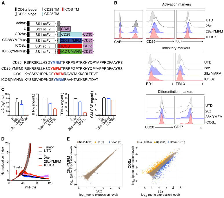
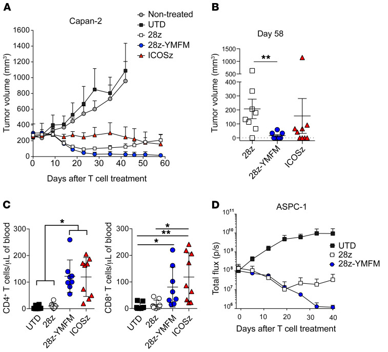
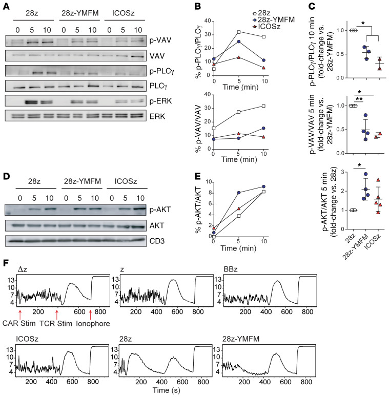
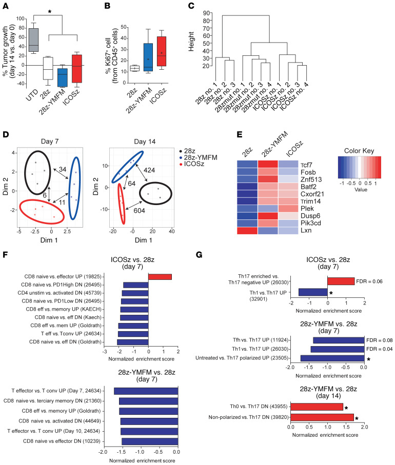
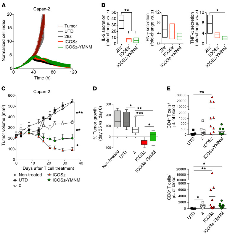
Chimeric antigen receptor-T (CAR-T) cell therapies can eliminate relapsed and refractory tumors, but the durability of antitumor activity requires in vivo persistence. Differential signaling through the CAR costimulatory domain can alter the T cell metabolism, memory differentiation, and influence long-term persistence. CAR-T cells costimulated with 4-1BB or ICOS persist in xenograft models but those constructed with CD28 exhibit rapid clearance. Here, we show that a single amino acid residue in CD28 drove T cell exhaustion and hindered the persistence of CD28-based CAR-T cells and changing this asparagine to phenylalanine (CD28-YMFM) promoted durable antitumor control. In addition, CD28-YMFM CAR-T cells exhibited reduced T cell differentiation and exhaustion as well as increased skewing toward Th17 cells. Reciprocal modification of ICOS-containing CAR-T cells abolished in vivo persistence and antitumor activity. This finding suggests modifications to the costimulatory domains of CAR-T cells can enable longer persistence and thereby improve antitumor response.

Keywords: Cancer immunotherapy; Immunology; T cells; Therapeutics.


Conflict of interest statement

Conflict of interest: ADP and SG are listed as the inventors on the patent application, WO2018140725A1, filed by the Trustees of the University of Pennsylvania and Novartis Ag.

Figures

Figure 1
Figure 1. Short-term signaling through 28z-YMFM CAR closely resembles CD28 signaling.
(A) Upper panel: Schematic representation of a panel of chimeric receptors that contain the SS1 scFv and differ in the intracellular domain. The CD28(YMFM)z mutant contains the CD28 transmembrane and intracellular domain with a point mutation in the YMNM motif (the asparagine [N] is mutated to phenylalanine [F]). The ICOS(YMNM)z mutant contains the ICOS transmembrane and intracellular domains with a point mutation in the YMFM motif [the phenylalanine [F] is mutated to an asparagine [N]). Lower panel: The full amino acid sequences for CD28 and ICOS intracellular domains and their corresponding mutants are shown. (B) Characterization of tonic signaling in CAR-T cells during primary expansion. Representative histograms showing the expression of the CAR protein and markers of activation, inhibition, and differentiation in CAR-T cells at 14 days after stimulation with anti-CD3/CD28 beads. (C) CAR-T cells were cocultured with mesothelin+ Capan-2 cells. Supernatants were obtained 24 hours later, and cytokine production was analyzed by Luminex. Representative of 2 donors. (D) A real-time cytotoxicity assay (xCELLigence) was used to evaluate the lysis of Capan-2 tumor cells when treated with CAR-T cells (E/T = 3:1) over a 120-hour period. (E) CAR-T cells from 2 different healthy donors were stimulated with immobilized recombinant mesothelin. Transcriptome analysis by RNA-sequencing was performed 6 days following antigen recognition. Scatter plots show the gene expression levels in 28z-YMFM or ICOSz CAR-T cells versus 28z CAR-T cells. The number of upregulated (orange) and downregulated (blue) genes is indicated.
Figure 2
Figure 2. The persistence of CD28-based CARs can be enhanced through mutation of the Grb2-interacting residue.
(A and B) NSG mice bearing s.c. Capan-2 tumors were treated 20 days after tumor implantation with 2 doses of control T cells (untransduced, UTD) or CAR-T cells (n = 8–9) on day 0 and day 15. (A) Tumor volume was analyzed at indicated time points. (B) Tumor volume on day 58 for individual animals is plotted. Error bars represent ± SEM (n = 8 tumors). **P < 0.01 by Kruskal-Wallis multiple-comparisons test. (C) The concentration of CD4+ and CD8+ T cells was determined in the blood of treated animals 30 days after T cell injection. Error bars represent ± SEM (n = 8–9). *P < 0.05; **P < 0.01 by Kruskal-Wallis multiple-comparisons test. (D) NSG mice bearing intraperitoneal CBG+ ASPC-1 tumors were treated 7 days after tumor implantation with 10 × 106 CAR-T cells. Bioluminescence was analyzed at the indicated time points (n = 4).
Figure 3
Figure 3. Signaling through a CD28-based CAR containing the ICOS YMFM motif shows enhanced AKT phosphorylation with reduced p-PLCγ, p-VAV, and calcium flux.
(AE) CAR-T cells were stimulated with magnetic beads coated with recombinant mesothelin. Cell lysates were obtained at 5 and 10 minutes following antigen encounter and phosphorylation levels for VAV, PLCγ, ERK, and AKT were analyzed by Western blot (A and D) and densitometry (B and E). Basal phosphorylation was evaluated without stimulation (minute 0). (C) T cells from 2 to 5 different healthy donors were stimulated as in AE, and AKT, VAV, and PLCγ phosphorylation was analyzed by densitometry. The mean ± SD from 4 independent experiments is shown. *P < 0.05; **P < 0.01 by 1-way ANOVA with Tukey’s post hoc test. (F) Calcium influx was measured in CAR-T cells at baseline for 30 seconds, and then after stimulation with mesothelin-coated magnetic beads for 6 minutes, followed by stimulation with biotinylated OKT3 and avidin for 6 minutes, and treated with ionomycin. The mean Indo-1 ratio of violet/blue fluorescence emission is displayed on the y axis and the time of sample collection in seconds is displayed on the x axis. Representative of 3 different experiments using 3 different normal donors.
Figure 4
Figure 4. In vivo long-term signaling through 28z-YMFM CAR is associated with a transcriptional profile that resembles ICOS signaling.
NSG mice bearing s.c. Capan-2 tumors were treated with CAR-T cells and tumor growth was monitored. T cells were isolated from tumors on day 7 and 14 after treatment for analysis. (A) Box plots of the change in tumor volume on day 14 versus baseline (n = 4–5). *P < 0.05 by 1-way ANOVA with Tukey’s post hoc test. (B) The expression of CD45 and Ki67 on T cells isolated on day 14 after treatment was analyzed by flow cytometry. For A and B, box plots show median (line), mean (plus symbol), and 25th to 75th percentile (box). The end of the whiskers represents the minimum and the maximum of all of the data. Error bars represent ± SEM (n = 4–5). P > 0.05. (C) Dendrogram showing the relatedness of gene expression patterns for individual animals. (D) MDS analysis of differentially expressed genes between the 3 sets of CAR-T cells isolated from Capan-2 xenograft tumors (n = 3–4) on day 7 and day 14. The number of differentially expressed genes (FDR < 0.05, FC < 2 or FC > 2) between a pair of CARs is indicated with pair-connecting arrows. (E) Heatmap showing differential expression of genes associated with T cell differentiation and exhaustion. Genes reported to be preferentially expressed in early stages of T cell differentiation (Tcf7, Fosb, Znf513, Batf2, Cxorf2, Trim14) and genes upregulated in effector cells (Lxn) or downregulated in effector or exhausted T cells (Dusp6 and Pik3cd) are plotted. (F and G) Normalized enrichment scores of selected up- or downregulated gene sets associated with T cell differentiation (F) or Th17 polarization (G) as determined by GSEA using MSigDB C7 gene ontology sets. *P < 0.05. For all pathways, FDR q ≤ 0.02 (unless otherwise indicated). Reference numbers for immunology-related gene lists from MSigDB C7 are indicated in parentheses.
Figure 5
Figure 5. Mutation of the YMFM motif in the ICOS signaling domain to YMNM (CD28 motif) impairs antitumor effect and T cell persistence.
(A) A real-time cytotoxicity assay (xCELLigence) was used to evaluate the lysis of Capan-2 tumor cells when treated with CAR-T cells (E/T = 3:1) over a 120-hour period. (B) CAR-T cells were cocultured with mesothelin+ Capan-2 cells. Supernatants were obtained 24 hours later, and cytokine production was analyzed by ELISA. Fold-change cytokine production in second-generation versus first-generation (z) CAR-T cells was analyzed in 3 to 4 healthy donors. P values were calculated by 1-way ANOVA with Tukey’s post hoc test. (CE) NSG mice bearing s.c. Capan-2 tumors were treated 30 days after tumor implantation with 2 doses of CAR-T cells (n = 6–9). (C) Tumor volume was analyzed at indicated time points. Statistical analysis was performed by 2-way ANOVA followed by Dunnett’s multiple-comparisons test. (D) Box plots of the change in tumor volume on day 35 versus baseline (n = 6–9). Box plots show median (line) and 25th to 75th percentile (box). The end of the whiskers represents the minimum and the maximum of all of the data. P values were calculated with 2-tailed Mann-Whitney tests. (E) The concentration of CD4+ and CD8+ T cells was determined in the blood of treated animals 36 days after T cell injection. P values were calculated by Kruskal-Wallis multiple-comparisons test. *P < 0.05; **P < 0.01; ***P < 0.001.

Comment in

References

    1. Maude SL, et al. Tisagenlecleucel in children and young adults with B-cell lymphoblastic leukemia. N Engl J Med. 2018;378(5):439–448. doi: 10.1056/NEJMoa1709866. - DOI - PMC - PubMed
    1. Neelapu SS, et al. Axicabtagene ciloleucel CAR T-cell therapy in refractory large B-cell lymphoma. N Engl J Med. 2017;377(26):2531–2544. doi: 10.1056/NEJMoa1707447. - DOI - PMC - PubMed
    1. Schuster SJ, et al. Tisagenlecleucel in adult relapsed or refractory diffuse large B-cell lymphoma. N Engl J Med. 2019;380(1):45–56. doi: 10.1056/NEJMoa1804980. - DOI - PubMed
    1. Brown CE, et al. Regression of glioblastoma after chimeric antigen receptor T-cell therapy. N Engl J Med. 2016;375(26):2561–2569. doi: 10.1056/NEJMoa1610497. - DOI - PMC - PubMed
    1. Louis CU, et al. Antitumor activity and long-term fate of chimeric antigen receptor-positive T cells in patients with neuroblastoma. Blood. 2011;118(23):6050–6056. doi: 10.1182/blood-2011-05-354449. - DOI - PMC - PubMed

Publication types

MeSH terms

Substances