. 2020 Mar 6:19:1186-1197.
doi: 10.1016/j.omtn.2020.01.007. Epub 2020 Jan 18.

Transplantation of hMSCs Genome Edited with LEF1 Improves Cardio-Protective Effects in Myocardial Infarction

Affiliations

Transplantation of hMSCs Genome Edited with LEF1 Improves Cardio-Protective Effects in Myocardial Infarction

Hyun-Min Cho et al. Mol Ther Nucleic Acids. .

Abstract

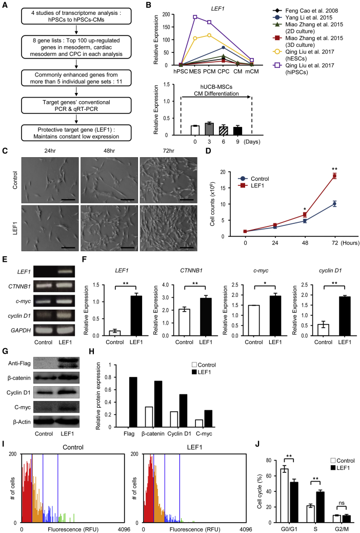
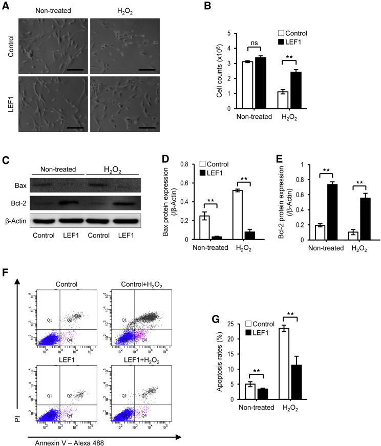
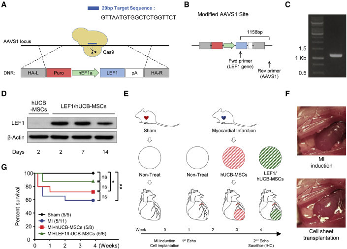
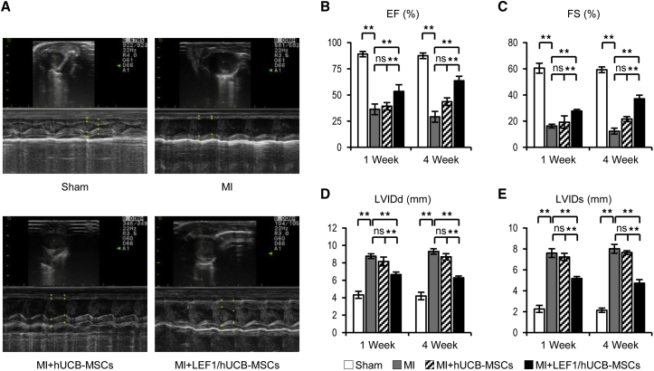
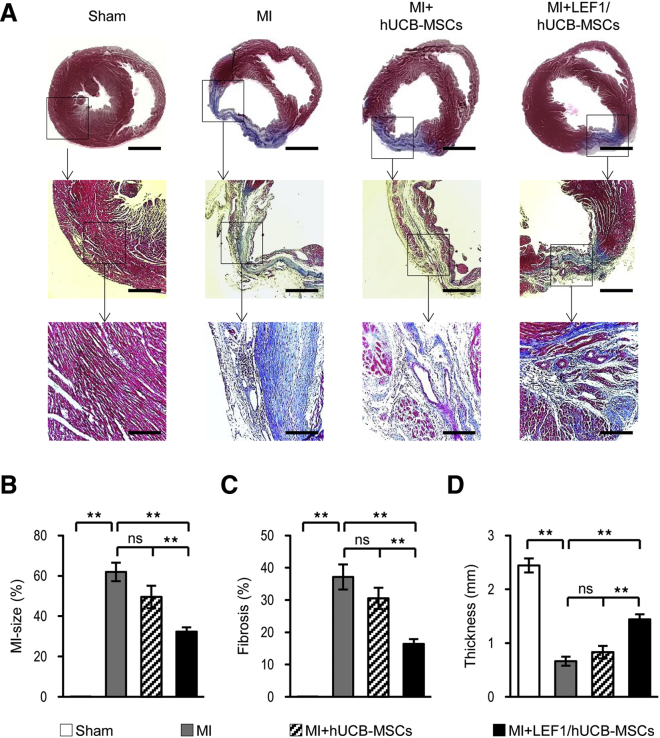
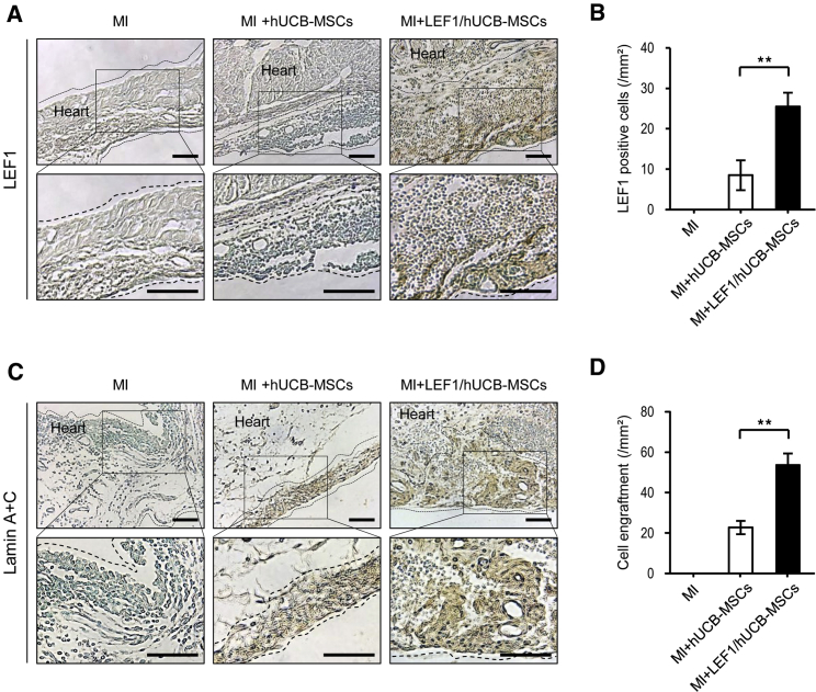
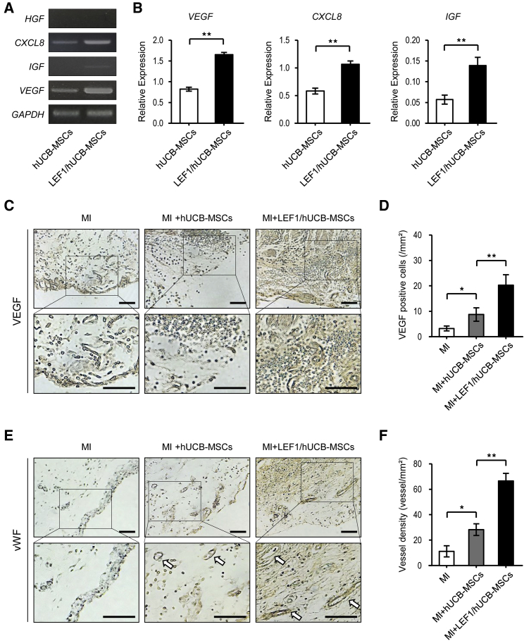
Stem cell-based therapy is one of the most attractive approaches to ischemic heart diseases, such as myocardial infarction (MI). We evaluated the cardio-protective effects of the human umbilical cord blood-derived mesenchymal stem cells (hUCB-MSCs) stably expressing lymphoid enhancer-binding factor 1 (LEF1; LEF1/hUCB-MSCs) in a rat model of MI. LEF1 overexpression in hUCB-MSCs promoted cell-proliferation and anti-apoptotic effects in hypoxic conditions. For the application of its therapeutic effects in vivo, the LEF1 gene was introduced into an adeno-associated virus integration site 1 (AAVS1) locus, known as a safe harbor site on chromosome 19 by CRISPR/Cas9-mediated gene integration in hUCB-MSCs. Transplantation of LEF1/hUCB-MSCs onto the infarction region in the rat model significantly improved overall survival. The cardio-protective effect of LEF1/hUCB-MSCs was proven by echocardiogram parameters, including greatly improved left-ventricle ejection fraction (EF) and fractional shortening (FS). Moreover, histology and immunohistochemistry successfully presented reduced MI region and fibrosis by LEF1/hUCB-MSCs. We found that these overall positive effects of LEF1/hUCB-MSCs are attributed by increased proliferation and survival of stem cells in oxidative stress conditions and by the secretion of various growth factors by LEF1. In conclusion, this study suggests that the stem cell-based therapy, conjugated with genome editing of transcription factor LEF1, which promotes cell survival, could be an effective therapeutic strategy for cardiovascular disease.

Keywords: CRISPR/Cas9; LEF1; cardio-protection; growth factors; hUCB-MSCs; myocardial infarction.


Figures

Figure 1
Figure 1
Selection Process of Therapeutic Target Gene LEF1 and Cell-Proliferation Effects of LEF1 in hUCB-MSCs (A) Schematic depiction of the overall workflow. (B) Comparison of LEF1 gene expression with in silico literature surveys and qRT-PCR data. (C) Representative images from phase-contrast microscopy at 24, 48, and 72 h after LEF1 transfection of hUCB-MSCs. Scale bars, 100 μm. Control (Ctrl): no DNA; LEF1: LEF1:pDC3.1. (D) hUCB-MSCs (1.5 × 105 cells), treated with no DNA or LEF1:pDC3.1, were seeded, and the increased number of cells was counted at the 24-, 48-, and 72-h time points. *p < 0.05 and **p < 0.01. (E) Conventional PCR for the Wnt pathway and cell-cycle-related genes. (F) Significant difference in gene-expression levels was confirmed by real-time PCR. Relative expression to GAPDH was calculated by the ΔΔ CT method. **p < 0.01. (G) Western blot analysis confirmed the increased expression in protein level under LEF1 overexpression. (H) Densitometry showed relative protein expression to β-actin level. (I) Cell-cycle analysis was performed by automated fluorescence cell counting in hUCB-MSCs differentially treated with no DNA or LEF1:pDC3.1. Colors indicate different stages; red: G0/G1 phase; yellow: S phase; blue: G2/M phase. (J) The histogram for the cell-cycle distribution after transfection of LEF1 and scrambled control DNA. **p < 0.01 compared to control.
Figure 2
Figure 2
LEF1 Overexpression Protects hUCB-MSCs from Oxidative Stress-Induced Apoptosis (A) Microscopy demonstrated that oxidative stress-caused cell death was attenuated by LEF1 overexpression. Scale bars, 100 μm. (B) Quantification of reduced cell death in LEF1 overexpression under oxidative stress condition. **p < 0.01. (C) Drastic increase in Bcl-2 and decrease in Bax expression in LEF1-overexpressing hUCB-MSCs were observed by western blot. (D and E) Image analysis confirmed the significant changes of Bax (D) and Bcl-2 (E) expression in LEF1-overexpressing hUCB-MSCs. (F) Fluorescence-activated cell sorting (FACS) analysis using PI and Annexin V successfully presented increased apoptotic cell populations (Q2) under H2O2 treatment in hUCB-MSCs (top right), but less cells were dead in LEF1-expressing hUCB-MSCs (bottom right). (G) Reduced apoptosis triggered by H2O2 in LEF1 transfection. **p < 0.01.
Figure 3
Figure 3
Experimental Strategy of the Therapeutic hUCB-MSC Transplantation System (A) Schematic diagram of the LEF1 gene-integration procedure by CRISPR/Cas9-mediated knockin to the AAVS1 site of hUCB-MSCs. 20-nt-length single-guide RNA (sgRNA) target sequence on the AAVS1 locus; LEF1 introduction cassette flanked by homologous arm (HA) left (HA-L) and right (HA-R) were indicated. (B) The architecture of donor DNA in the AAVS1 locus. Arrows: primers designed for the detection of successful homologous recombination. (C) Confirmation of the correct integration of the LEF1 cassette into the AAVS1 locus using PCR (1,158 expected size). (D) Western blot analysis showing stable expression of LEF1 protein from LEF1/hUCB-MSCs for 2 weeks. (E) Schematic illustration of the therapeutic procedure with cell-sheet transplantation of hUCB-MSCs and LEF1/hUCB-MSCs in an MI model. Of note, the cell-sheet transplantation was performed with induction surgery of MI on day 0, and echocardiography measurements were performed prior to MI surgery, as well as 1 week and 4 weeks after transplantation. The four groups are Sham: surgery without MI; MI: MI alone; MI + hUCB-MSCs: MI treated with hUCB-MSCs; and MI + LEF1/hUCB-MSCs: MI treated with LEF1/hUCB-MSCs. (F) Representative images depicting before and after MI induction and stem cell transplantation using the UpCell system. (G) Survival curves of the experimental groups. The survival rate was significantly enhanced in the LEF1/hUCB-MSC group (n = 5–11). *p < 0.05; **p < 0.01; ns, not significant.
Figure 4
Figure 4
Transplantation of LEF1/hUCB-MSCs Recovered Cardiac Function in MI Rat (A) Representative images of echocardiography showed successively improved cardiac function in MI, MI + hUCB-MSCs, and MI + LEF1/hUCB-MSCs at 4 weeks postsurgery. (B–E) Four values representing cardiac function (EF in B; FS in C; LVIDd in D; and LVIDs in E) were measured and compared in histograms. Significant improvements were detected in all MI + LEF1/hUCB-MSC groups.*p < 0.05, **p < 0.01; ns, not significant. Error bar: standard error. White bars, Sham; gray bars, MI; striped bars, MI + hUCB-MSCs; black bars, MI + LEF1/UCB-MSCs.
Figure 5
Figure 5
The LEF1/hUCB-MSC Transplantation Greatly Reduced MI Size and Fibrosis and Restored the LV Wall Thickness (A) Masson’s trichrome staining showed MI regions at 4 weeks after surgery. Scale bars: top row, 1 mm; middle row, 400 μm; bottom row, 200 μm. The red-stained region means the viable myocardium, and the blue-stained region means the fibrotic area. The large, fibrous region stained in blue is found in MI alone compared to MI + hUCB-MSCs and MI + LEF1/hUCB-MSCs. Serial magnification distinctively presented damaged heart in MI and enhanced heart-protective efficacy of MI + LEF1/hUCB-MSCs. (B–D) Quantification of infarct size (B), fibrosis region (C), and the wall thickness (D) of LV in each group (n = 5). *p < 0.05, **p < 0.01; ns, not significant.
Figure 6
Figure 6
Immunohistochemical Staining Confirmed the Cells Surviving and Expressing LEF1 from the Engrafted MI + LEF1/hUCB-MSCs (A) LEF1 was detected only from MI + LEF1/hUCB-MSCs. No rat heart cells and hUCB-MSCs expressed LEF1. Scale bars, 100 μm. (B) LEF1-positive cells in the heart tissue were quantitatively measured (n = 5). (C) No lamin signal was detected in the MI-alone group. A thin layer was found in the group of MI + hUCB-MSCs, and a thicker layer was stained from MI + LEF1/hUCB-MSC-engrafted MI. Scale bars, 100 μm. (D) Human cell engraftment was quantitatively measured (n = 5). **p < 0.01.
Figure 7
Figure 7
LEF1 Triggered Therapeutic Gene (VEGF, IL-8, IGF) Expressions and Enhanced Angiogenesis in MI + LEF1/hUCB-MSCs (A) Expression of growth factors HGF, CXCL8, IGF, and VEGF was measured by conventional PCR. (B) Increased mRNA expression of growth factors in LEF1/hUCB-MSCs compared with hUCB-MSCs. Real-time PCR demonstrated that the relative gene expressions of VEGF, CXCL8, and IGF to GAPDH were increased in LEF1/hUCB-MSCs. **p < 0.01. (C) Immunohistochemical staining for VEGF in three MI groups: MI alone, MI + hUCB-MSCs, and LEF1/MI + hUCB-MSCs. Anti-VEGF antibody faintly stained rat heart, but a stronger signal was detected in LEF1/hUCB-MSCs. Scale bars, 100 μm. (D) VEGF protein expression was measured in infarcted heart among different groups (n = 5). (E) Immunohistochemical staining for vWF in three MI groups: MI alone, MI + hUCB-MSCs, and LEF1/MI + hUCB-MSCs. The blood vessels are stained in brown (arrows). More vessels were observed in MI + LEF1/hUCB-MSCs. Scale bars, 100 μm. (F) Higher vascular densities were detected in the MI + LEF1/hUCB-MSC group than in the MI-alone and MI + hUCB-MSC groups (n = 5). *p < 0.05 and **p < 0.01.

References

    1. GBD 2016 Causes of Death Collaborators Global, regional, and national age-sex specific mortality for 264 causes of death, 1980–016: a systematic analysis for the Global Burden of Disease Study 2016. Lancet. 2017;390:1151–1210. - PMC - PubMed
    1. Jessup M., Brozena S. Heart failure. N. Engl. J. Med. 2003;348:2007–2018. - PubMed
    1. Neri M., Riezzo I., Pascale N., Pomara C., Turillazzi E. Ischemia/Reperfusion Injury following Acute Myocardial Infarction: A Critical Issue for Clinicians and Forensic Pathologists. Mediators Inflamm. 2017;2017:7018393. - PMC - PubMed
    1. Nagamura-Inoue T., He H. Umbilical cord-derived mesenchymal stem cells: Their advantages and potential clinical utility. World J. Stem Cells. 2014;6:195–202. - PMC - PubMed
    1. Amado L.C., Saliaris A.P., Schuleri K.H., St John M., Xie J.S., Cattaneo S., Durand D.J., Fitton T., Kuang J.Q., Stewart G. Cardiac repair with intramyocardial injection of allogeneic mesenchymal stem cells after myocardial infarction. Proc. Natl. Acad. Sci. USA. 2005;102:11474–11479. - PMC - PubMed

LinkOut - more resources