. 2020 Feb 13;12(2):436.
doi: 10.3390/polym12020436.

Thermosensitive Chitosan-Gelatin-Glycerol Phosphate Hydrogels as Collagenase Carrier for Tendon-Bone Healing in a Rabbit Model

Affiliations

Thermosensitive Chitosan-Gelatin-Glycerol Phosphate Hydrogels as Collagenase Carrier for Tendon-Bone Healing in a Rabbit Model

Yu-Min Huang et al. Polymers (Basel). .

Abstract

Healing of an anterior cruciate ligament graft in bone tunnel yields weaker fibrous scar tissue, which may prolong an already prolonged healing process within the tendon-bone interface. In this study, gelatin molecules were added to thermosensitive chitosan/β-glycerol phosphate disodium salt hydrogels to form chitosan/gelatin/β-glycerol phosphate (C/G/GP) hydrogels, which were applied to 0.1 mg/mL collagenase carrier in the tendon-bone junction. New Zealand white rabbit's long digital extensor tendon was detached and translated into a 2.5-mm diameter tibial plateau tunnel. Thirty-six rabbits underwent bilateral surgery and hydrogel injection treatment with and without collagenase. Histological analyses revealed early healing and more bone formation at the tendon-bone interface after collagenase partial digestion. The area of metachromasia significantly increased in both 4-week and 8-week groups after collagenase treatment (p < 0.01). Micro computed tomography showed a significant increase in total bone volume and bone volume/tissue volume in the 8 weeks after collagenase treatment, compared with the control group. Load-to-failure was significantly higher in the treated group at 8 weeks (23.8 ± 8.13 N vs 14.3 ± 3.9 N; p = 0.008). Treatment with collagenase digestion resulted in a 66% increase in pull-out strength. In conclusion, injection of C/G/GP hydrogel with collagenase improves tendon-to-bone healing in a rabbit model.

Keywords: collagenase digestion; hydrogel; rabbit model; tendon–bone healing.


Conflict of interest statement

The authors declare no conflict of interest.

Figures

Figure 1
Figure 1
Animal study design.
Figure 2
Figure 2
Cytotoxic of chitosan/gelatin/β-glycerol phosphate (C/G/GP) with collagenase to tenocyte. Figure 2a shows water soluble tetrazolium salt-1 (WST-1) assay (n = 5, p > 0.05) under different collagenase concentration and Figure 2b shows lactate dehydrogenase assay (n = 5, p > 0.05).
Figure 3
Figure 3
Release profile of the collagenases loaded in C/G/GP hydrogel. The release profile was evaluated by incubation C/G/GP hydrogel in PBS with collagenase concentration of 0.1 mg/mL. The error bars designed the standard deviation.
Figure 4
Figure 4
Necropsy examination. Photography at necropsy showed bone-tunnel healing in a rabbit model. A bony defect was identified in the control group at 4 weeks (a) and 8 weeks (b). Better bony ingrowth with a decrease in tunnel diameter was seen in the study group at 4 weeks (c) and 8 weeks (d) (scale bar: 1 mm).
Figure 5
Figure 5
Four-week specimens histology. Histology of a tendon (T)-to-bone (B) interface (IF) in a 4-week specimen in the control group (a) and the study group (b). A wider interface with dense fibrovascular tissue formation in the study group was found. Sharpey fiber-like tissue was more observed 4 weeks after collagenase treatment (c,d) (scale bar: 100 μm).
Figure 6
Figure 6
Eight-week specimens histology. Wider dense interface tissue between the tendon (T) and bone (B) in the 8-week study group (a,b). New bone formation with incorporation into the tendon graft is illustrated (c,d). The tendon graft was replaced by new bone formation after collagenase treatment. (scale bar: 100 μm).
Figure 7
Figure 7
Metachromacia. The area of metachromcia was significantly increased in both 4-week and 8-week groups after collagenase treatment (p < 0.01). The error bars designed the standard deviation.
Figure 8
Figure 8
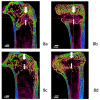
Microcomputed tomography (Micro-CT) examination showed bone formation. Micro-CT shows tendon graft and bone tunnel in an 8-week specimen from the control group. (a,b) Bone tunnel obliteration with new bone ingrowth was identified in sagittal and coronal views after enzyme treatment (c,d). Prominent bone ingrowth was observed in both tunnel orifices. Line arrow marks the tendon graft, and block arrows indicate the bone tunnel. (scale bar: 1 mm).
Figure 9
Figure 9
Micro-CT evaluation. Total bone volume (mm3 ± SD) determined by micro-CT. The region of interest was measured following the long axis of the bone tunnel. (a) There was significantly more new bone formation at 8 weeks after collagenase treatment. Total bone volume/tissue volume was illustrated, and the ratio was significant 8 weeks after enzyme treatment. (b) p < 0.05, n = 6. The error bars designed the standard deviation.
Figure 10
Figure 10
Biomechanical assessment. Original load-deformation curve in the 8-week enzyme-treated specimen. (a) Significantly higher tendon ultimate load-to-failure and stress were found in the enzyme treatment group than in the control group at 8 weeks. * p < 0.05, n = 8. (b,c) The error bars designed the standard deviation.
Figure 10
Figure 10
Biomechanical assessment. Original load-deformation curve in the 8-week enzyme-treated specimen. (a) Significantly higher tendon ultimate load-to-failure and stress were found in the enzyme treatment group than in the control group at 8 weeks. * p < 0.05, n = 8. (b,c) The error bars designed the standard deviation.

References

    1. Frank C.B., Jackson D.W. Current concepts review—The science of reconstruction of the anterior cruciate ligament. J. Bone Jt. Surg. 1997;79:1556–1576. doi: 10.2106/00004623-199710000-00014. - DOI - PubMed
    1. Freedman K.B., D’Amato M.J., Nedeff D.D., Kaz A., Bach B.R. Arthroscopic anterior cruciate ligament reconstruction: A metaanalysis comparing patellar tendon and hamstring tendon autografts. Am. J. Sports Med. 2003;31:2–11. doi: 10.1177/03635465030310011501. - DOI - PubMed
    1. Poehling-Monaghan K.L., Salem H., Ross K.E., Secrist E., Ciccotti M.C., Tjoumakaris F., Ciccotti M.G., Freedman K.B. Long-term outcomes in anterior cruciate ligament reconstruction: A systematic review of patellar tendon versus hamstring autografts. Orthop. J. Sports Med. 2017;5 doi: 10.1177/2325967117709735. - DOI - PMC - PubMed
    1. Gifstad T., Foss O.A., Engebretsen L., Lind M., Forssblad M., Albrektsen G., Drogset J.O. Lower risk of revision with patellar tendon autografts compared with hamstring autografts: A registry study based on 45,998 primary ACL reconstructions in Scandinavia. Am. J. Sports Med. 2014;42:2319–2328. doi: 10.1177/0363546514548164. - DOI - PubMed
    1. Samuelsen B.T., Webster K.E., Johnson N.R., Hewett T.E., Krych A.J. Hamstring Autograft versus Patellar Tendon Autograft for ACL Reconstruction: Is There a Difference in Graft Failure Rate? A Meta-analysis of 47,613 Patients. Clin. Orthop. Relat. Res. 2017;475:2459–2468. doi: 10.1007/s11999-017-5278-9. - DOI - PMC - PubMed

LinkOut - more resources