

BSCN: bidirectional symmetric cascade network for retinal vessel segmentation
- PMID: 32070306
- PMCID: PMC7029442
- DOI: 10.1186/s12880-020-0412-7
BSCN: bidirectional symmetric cascade network for retinal vessel segmentation
Abstract
Background: Retinal blood vessel segmentation has an important guiding significance for the analysis and diagnosis of cardiovascular diseases such as hypertension and diabetes. But the traditional manual method of retinal blood vessel segmentation is not only time-consuming and laborious but also cannot guarantee the accuracy and efficiency of diagnosis. Therefore, it is especially significant to create a computer-aided method of automatic and accurate retinal vessel segmentation.
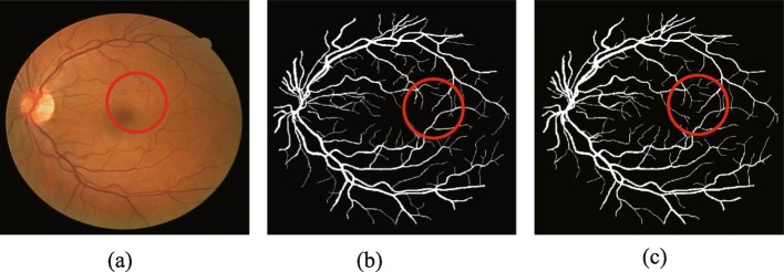
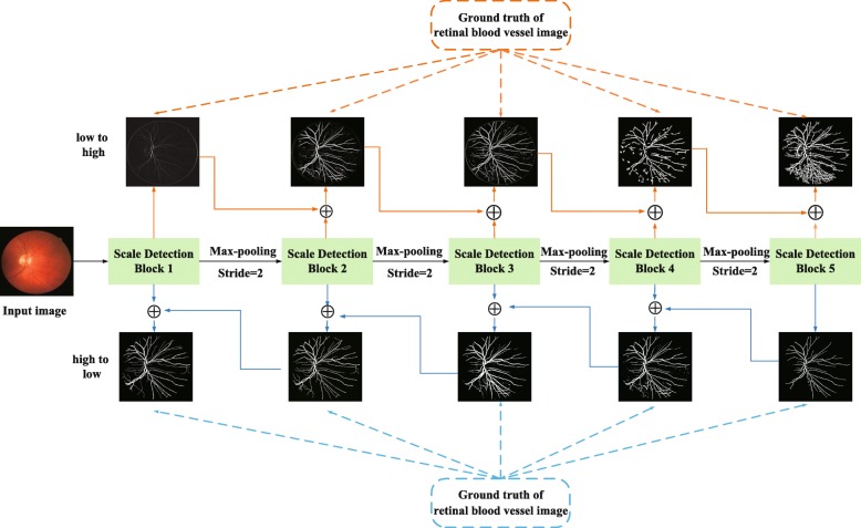
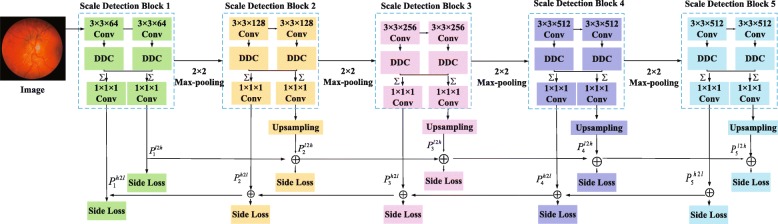
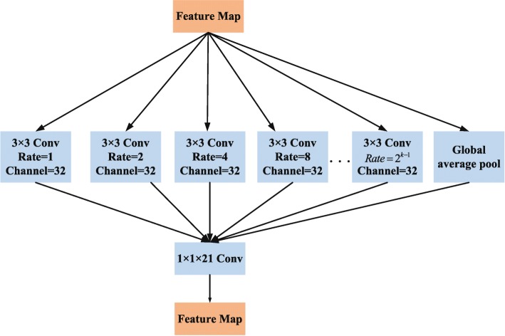
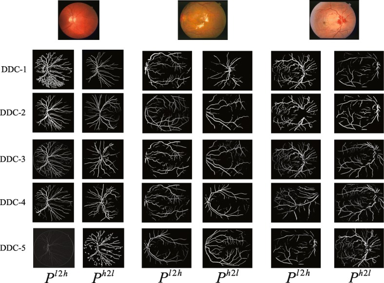
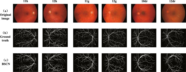
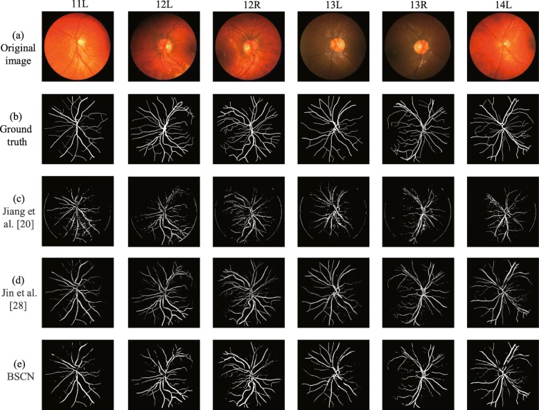
Methods: In order to extract the blood vessels' contours of different diameters to realize fine segmentation of retinal vessels, we propose a Bidirectional Symmetric Cascade Network (BSCN) where each layer is supervised by vessel contour labels of specific diameter scale instead of using one general ground truth to train different network layers. In addition, to increase the multi-scale feature representation of retinal blood vessels, we propose the Dense Dilated Convolution Module (DDCM), which extracts retinal vessel features of different diameters by adjusting the dilation rate in the dilated convolution branches and generates two blood vessel contour prediction results by two directions respectively. All dense dilated convolution module outputs are fused to obtain the final vessel segmentation results.
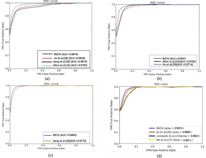
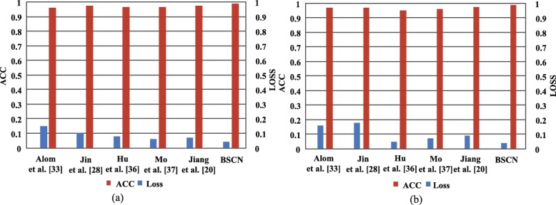
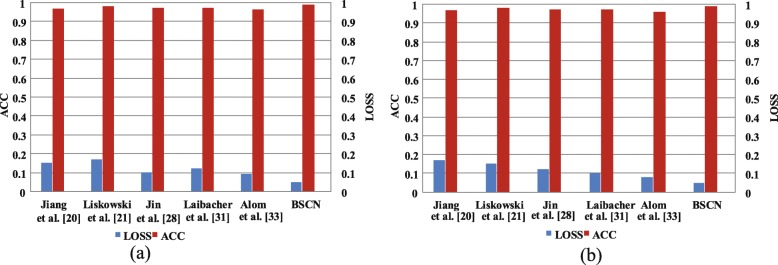
Results: We experimented the three datasets of DRIVE, STARE, HRF and CHASE_DB1, and the proposed method reaches accuracy of 0.9846/0.9872/0.9856/0.9889 and AUC of 0.9874/0.9941/0.9882/0.9874 on DRIVE, STARE, HRF and CHASE_DB1.
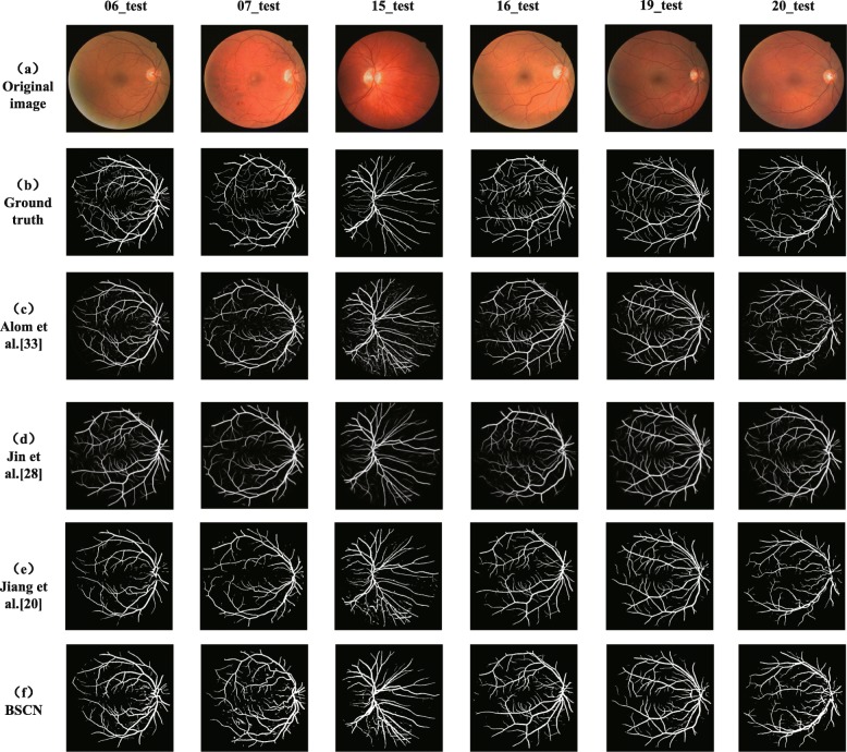
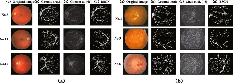
Conclusions: The experimental results show that compared with the state-of-art methods, the proposed method has strong robustness, it not only avoids the adverse interference of the lesion background but also detects the tiny blood vessels at the intersection accurately.
Keywords: Bidirectional symmetric cascade network; Dense dilated convolution; Retinal vessel segmentation; Scale detection; Specific diameter scale.
Conflict of interest statement
The authors declare that they have no competing interests.
Figures
Similar articles
-
Retinal blood vessel extraction employing effective image features and combination of supervised and unsupervised machine learning methods.Artif Intell Med. 2019 Apr;95:1-15. doi: 10.1016/j.artmed.2019.03.001. Epub 2019 Mar 2. Artif Intell Med. 2019. PMID: 30904129
-
Densely connected U-Net retinal vessel segmentation algorithm based on multi-scale feature convolution extraction.Med Phys. 2021 Jul;48(7):3827-3841. doi: 10.1002/mp.14944. Epub 2021 Jun 16. Med Phys. 2021. PMID: 34028030
-
Retinal blood vessel segmentation using fully convolutional network with transfer learning.Comput Med Imaging Graph. 2018 Sep;68:1-15. doi: 10.1016/j.compmedimag.2018.04.005. Epub 2018 Apr 26. Comput Med Imaging Graph. 2018. PMID: 29775951
-
Retinal Vessel Segmentation, a Review of Classic and Deep Methods.Ann Biomed Eng. 2022 Oct;50(10):1292-1314. doi: 10.1007/s10439-022-03058-0. Epub 2022 Aug 25. Ann Biomed Eng. 2022. PMID: 36008569 Review.
-
Automated techniques for blood vessels segmentation through fundus retinal images: A review.Microsc Res Tech. 2019 Feb;82(2):153-170. doi: 10.1002/jemt.23172. Epub 2019 Jan 5. Microsc Res Tech. 2019. PMID: 30614150 Review.
Cited by
-
State-of-the-art retinal vessel segmentation with minimalistic models.Sci Rep. 2022 Apr 13;12(1):6174. doi: 10.1038/s41598-022-09675-y. Sci Rep. 2022. PMID: 35418576 Free PMC article.
-
Diabetic and Hypertensive Retinopathy Screening in Fundus Images Using Artificially Intelligent Shallow Architectures.J Pers Med. 2021 Dec 23;12(1):7. doi: 10.3390/jpm12010007. J Pers Med. 2021. PMID: 35055322 Free PMC article.
-
Practical utility of liver segmentation methods in clinical surgeries and interventions.BMC Med Imaging. 2022 May 24;22(1):97. doi: 10.1186/s12880-022-00825-2. BMC Med Imaging. 2022. PMID: 35610600 Free PMC article.
-
Retinal Vessel Automatic Segmentation Using SegNet.Comput Math Methods Med. 2022 Mar 26;2022:3117455. doi: 10.1155/2022/3117455. eCollection 2022. Comput Math Methods Med. 2022. Retraction in: Comput Math Methods Med. 2023 Oct 18;2023:9828704. doi: 10.1155/2023/9828704. PMID: 35378728 Free PMC article. Retracted.
-
Recent developments on computer aided systems for diagnosis of diabetic retinopathy: a review.Multimed Tools Appl. 2023;82(10):14471-14525. doi: 10.1007/s11042-022-13841-9. Epub 2022 Sep 24. Multimed Tools Appl. 2023. PMID: 36185322 Free PMC article.
References
-
- Irshad S, Akram MU. Classification of retinal vessels into arteries and veins for detection of hypertensive retinopathy. In: Lewin RA, editor. Proceedings of 7th Cairo International Biomedical Engineering Conference 11-13 December 2014. Giza: IEEE; 2014.
Publication types
MeSH terms
LinkOut - more resources
Full Text Sources
