. 2020 Feb 18;13(1):22.
doi: 10.1186/s13041-020-0555-z.

Upregulation of Beta4 subunit of BKCa channels in the anterior cingulate cortex contributes to mechanical allodynia associated anxiety-like behaviors

Affiliations

Upregulation of Beta4 subunit of BKCa channels in the anterior cingulate cortex contributes to mechanical allodynia associated anxiety-like behaviors

Huan Zhao et al. Mol Brain. .

Abstract

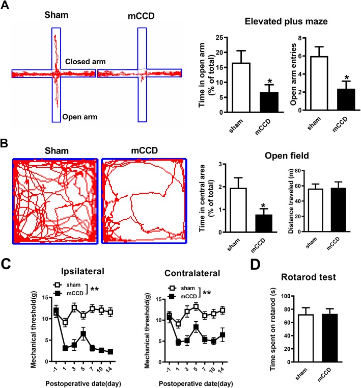
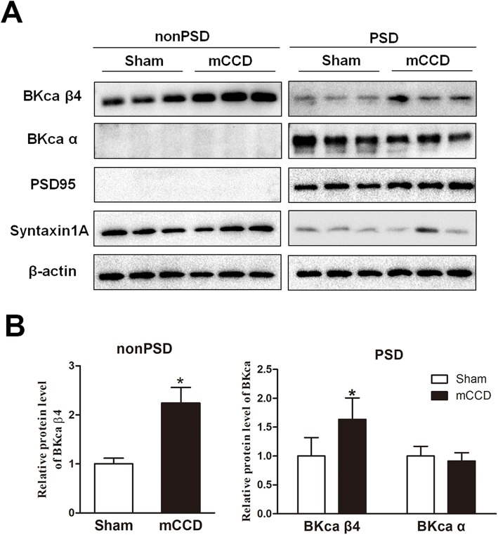
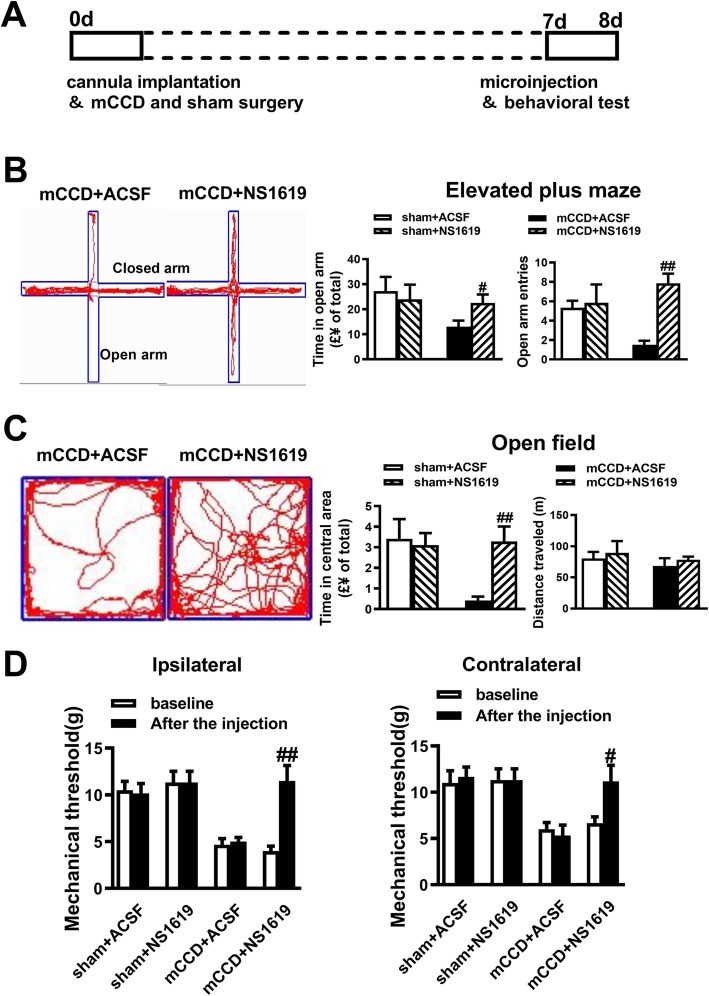
The anterior cingulate cortex (ACC) serves as a critical hub for the anxiety and pain perception. The large-conductance Ca2+-activated potassium channels, or BKCa channels, are ubiquitously expressed throughout the central nervous system including the cingulate cortex. However, what changes of cortical BKCa channels undergo in the ACC remains unknown in pain-related anxiety. In the present study, a significant upregulation of synaptic and non-synaptic BKCa channel accessory β4 subunits in the ACC was accompanied with pain-associated anxiety-like behaviors in the chronic compression of multiple dorsal root ganglia (mCCD) of the rat. NS1619, an opener of BKCa channels, significantly rescued the alteration of fAHP and AP duration of ACC pyramidal neurons in mCCD rats. The mRNA expression of BKCa β4 subunits was extremely upregulated in the ACC after mCCD with the increased amount of both synaptic and non-synaptic BKCa β4 subunit protein. Meanwhile, NS1619 reversed the enhanced AMPA receptor-mediated spontaneous excitatory postsynaptic current (sEPSC) frequency and the attenuated PPR of ACC neurons in mCCD rats. Local activation of BKCa channels in the ACC reversed mechanical allodynia and anxiety-like behaviors. These results suggest that the upregulation of postsynaptic and presynaptic BKCa β4 subunit may contribute to neuronal hyperexcitability and the enhanced synaptic transmission in the ACC in neuropathic pain state, and then may result in anxiety-like behavior induced by neuropathic pain.

Keywords: Anterior cingulate cortex; Anxiety; Excitability; Large-conductance Ca2+-activated potassium channel; Synaptic transmission.


Conflict of interest statement

The authors declare that they have no competing interests.

Figures

Fig. 1
Fig. 1
Enhanced anxiety in neuropathic pain rats following mCCD. a, Representative traces show the movement of sham and mCCD rats in elevated plus maze for 5 mins, mCCD rats (n = 13) spent less time in the open arms of elevated plus maze and showed a decrease in open arm entries compared with sham rats (n = 13). b, Representative traces show the movement of sham and mCCD rats in the open field test over a period of 15 min, mCCD rats (n = 13) spent significantly less time in the central zone in the open field test compared with sham rats (n = 13). Total distance travelled in the open field test was not changed compared with sham rats. c, The time course of ipsilateral and contralateral hind paw mechanical withdrawal thresholds after mCCD (sham group: n = 11, mCCD group: n = 8). The ipsilateral and contralateral hind paw withdrawal thresholds were significantly decreased below baseline on the 1st postoperative day and reached their lowest peak during the 7th to 14th postoperative day. d, Motor performances on the accelerating rotarod of mCCD rats (mCCD: n = 7, sham: n = 7). *p < 0.05, **p < 0.01 compared with that of sham group
Fig. 2
Fig. 2
Elevated excitability of pyramidal neurons in the ACC in mCCD rats. a, Representative traces showing the firing property in the neurons from ACC of sham and mCCD rats response to a 300 pA depolarizing current injection (500 ms). b, Representative recording of the first spike in the neurons from sham and mCCD slices. c, The amplitude of fAHP of the first three spikes in a train. d-i, The duration (d), half width (e), decay time (f), dacay slope (g), rise slope (h), rise time (i) of the first spike of pyramidal neurons from mCCD and sham rats (mCCD: n = 24; sham: n = 35) *p < 0.05, **p < 0.01 compared with that of sham group
Fig. 3
Fig. 3
Effects of NS1619 and paxilline on the excitability of pyramidal neurons in the ACC following mCCD. a-b, Representative recording traces of the first spike in the neurons from sham and mCCD slices in the presence of paxilline or NS1619 respectively. c, Histograms show the amplitude of fAHP of the first three spikes from the sham group in the absence and presence of paxilline (10 μM) (sham, n = 30, sham+paxilline: n = 10), and from the mCCD group in the absence and presence of NS1619 (10 μM) (mCCD: n = 48, mCCD+NS1619: n = 17).d-f, Histograms show AP duration (d), AP half-width (e) and AP decay time (f) from the sham group in the absence and presence of paxilline (sham: n = 30, sham+paxilline: n = 10), and from the mCCD group in the absence and presence of NS1619 (mCCD: n = 48, mCCD+NS1619: n = 17). *p < 0.05, **p < 0.01compared with that of mCCD group
Fig. 4
Fig. 4
Decreased BKCa currents of ACC pyramidal neurons from mCCD rats. a, Voltage ranges from − 60 to + 40 mV with 10 mV increments, typical recordings of BKCa currents in pyramidal neurons from sham (left) and mCCD (right) rats, BKCa currents were isolated with paxilline (10 μM). b, The I-V relationship curves showed the differences in the ACC pyramidal neurons from mCCD (n = 12) and sham (n = 12) rats. c, BKCa current density at + 40 mV from voltage-clamp recordings of ACC pyramidal neurons from mCCD (n = 12) and sham (n = 12) rats. **p < 0.01 compared with that of sham group
Fig. 5
Fig. 5
Upregulation of BKCa β4 subunit protein and mRNA in the ACC during neuropathic pain associated anxiety-like behavior. a, The schematic diagram of the behavioral and biochemical experiments. b, Quantification of the mRNA levels of BKCa channel subunits in the ACC between mCCD (n = 7) and sham (n = 6) rats on postoperative day 7. c, Quantification of the mRNA levels of BKCa channels in the insular cortex between mCCD (n = 7) and sham (n = 6) rats on postoperative day 7. d, Quantification of the mRNA levels of BKCa β4 subunit in the ACC between mCCD (n = 6) and sham (n = 6) rats on postsurgical day 3, 7, 14. e, Representative Western blots for BKCa α and BKCa β4 subunit in the ACC obtained on postsurgical day 7. f, Quantification of the protein levels of α and β4 subunits in the ACC between mCCD (n = 3) and sham (n = 3) rats on postoperative day 7. *p < 0.05, **p < 0.01 compared with that of sham group
Fig. 6
Fig. 6
Postsynaptic and presynaptic upregulation of BKCa β4 subunit protein in the ACC during neuropathic pain associated anxiety-like behavior. a, Representative Western blots for PSD 95, syntaxin 1A, BKCa β4 and BKCa α subunits in the non-PSD and PSD membrane fractions of the ACC in sham and mCCD rats on postoperative day 14; b, BKCa β4 subunit was significantly enhanced in the PSD and non-PSD fractions of the ACC from mCCD (PSD fraction: 164 ± 37%, n = 6, *p < 0.05; non-PSD fraction: 224 ± 32%, n = 6, *p < 0.05) rats on postoperative day 14 compared with those in sham rats (PSD fraction: 100 ± 32%, n = 6; non-PSD fraction: 100 ± 12%, n = 6). BKCa α subunit showed no significant change in the PSD fraction between sham (100 ± 17%, n = 6) and mCCD (91.4 ± 14%, n = 6, p > 0.05) rats on postoperative day 14
Fig. 7
Fig. 7
NS1619 decreased the increased AMPA receptor-mediated sEPSC frequency of ACC neurons in mCCD rats. a, Representative sEPSCs recorded in ACC pyramidal neuron in slices from sham and mCCD rats at a holding potential of − 60 mV; b, Cumulative interevent interval (top) and amplitude (bottom) histograms of sEPSCs; f, Summary plots of mean sEPSC peak frequency and amplitude (n = 13 neurons for sham and n = 16 neurons for mCCD). c, Representative traces show AMPA receptor-mediated sEPSCs in the presence of paxilline (10 μM) in sham rats and those in the presence of NS1619 (10 μM) in mCCD rats; d, Cumulative interevent interval and amplitude histograms of sEPSCs in the presence of paxilline (10 μM) in sham rats; e, Cumulative interevent interval and amplitude histograms of sEPSCs of ACC neurons in the presence of NS1619 in mCCD rats; f, Summary plots of sEPSC peak frequency and amplitude in the presence of paxilline (10 μM) in sham rats and those in the presence of NS1619 (10 μM) in mCCD rats(n = 13 neurons for sham and n = 16 neurons for mCCD). *p < 0.05, compared to the value of sham group, #p < 0.05, ##p < 0.01 compared with that of mCCD group
Fig. 8
Fig. 8
NS1619 enhanced the decreased PPR of ACC neurons in mCCD rats. a-b, Representative traces with an interval of 50 ms recorded in layer II/III of the ACC; a, PPR (the ratio of EPSC2 / EPSC1) were recorded at intervals of 50 ms from sham rats (n = 6 neurons in the absence or presence of paxilline (10 μM). b, PPR were recorded at intervals of 50 ms from mCCD rats (n = 8 neurons in the absence or presence of NS1619 (10 μM). c, Summary plots of PPR at intervals of 50 ms from sham and mCCD rats (n = 6 neurons for sham and n = 8 neurons for mCCD), and effects of paxilline and NS1619 on PPR of sham and mCCD rats respectively. *p < 0.05 compared with that of sham group, #p < 0.05 compared to that of mCCD group
Fig. 9
Fig. 9
Effects of activating BKCa channels in the ACC on neuropathic pain and anxiety-like behaviors. a, A schematic diagram of microinjection and the behavioral experiment. b, Bilateral microinjection of NS1619 (10 μM, 0.5 μL) into the ACC reversed the time in the open arms and the open arm entries in mCCD rats on day 7 after surgery. c, Rats infused with NS1619 spent more time in the central areas in open field test in mCCD rats compared with ACSF-treated animals. There was no significant difference in the total distance travelled within the open field for 15 min among mCCD and sham rats (sham + ACSF: n = 6, sham +NS1619: n = 6, mCCD + ACSF: n = 6, mCCD +NS1619: n = 6). d, The pain threshold of the injured hind paw (left) and contralateral feet (right), after bilateral microinjection of NS1619 into the ACC on day 7 after surgery. #p < 0.05, ##p < 0.01 compared with that of mCCD with ACSF group

Similar articles

Cited by

References

    1. Barthas F, Sellmeijer J, Hugel S, Waltisperger E, Barrot M, Yalcin I. The anterior cingulate cortex is a critical hub for pain-induced depression. Biol Psychiatry. 2015;77(3):236–245. doi: 10.1016/j.biopsych.2014.08.004. - DOI - PubMed
    1. Bliss TV, Collingridge GL, Kaang BK, Zhuo M. Synaptic plasticity in the anterior cingulate cortex in acute and chronic pain. Nat Rev Neurosci. 2016;17(8):485–496. doi: 10.1038/nrn.2016.68. - DOI - PubMed
    1. Zhuo M. Cortical excitation and chronic pain. Trends Neurosci. 2008;31(4):199–207. doi: 10.1016/j.tins.2008.01.003. - DOI - PubMed
    1. Zhuo M. Neural mechanisms underlying anxiety-chronic pain interactions. Trends Neurosci. 2016;39(3):136–145. doi: 10.1016/j.tins.2016.01.006. - DOI - PubMed
    1. Sun YG, Gao YJ, Zhao ZQ, Huang B, Yin J, Taylor GA, Chen ZF. Involvement of P311 in the affective, but not in the sensory component of pain. Molecular pain. 2008;4:23. doi: 10.1186/1744-8069-4-23. - DOI - PMC - PubMed

Publication types

MeSH terms