

Hip Pain in Children
- PMID: 32070474
- PMCID: PMC7054595
- DOI: 10.3238/arztebl.2020.0072
Hip Pain in Children
Abstract
Background: Atraumatic hip pain in children is one of the most common symptoms with which pediatricians, orthopedists, and general practitioners are confronted, with an incidence of 148 cases per 100 000 persons per year.
Methods: This article is based on publications up to April 2019 that were retrieved by a selective search in the PubMed data- base, including case reports and reviews.
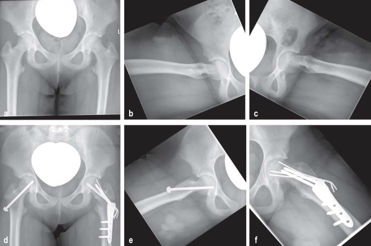
Results: Infants with fever often have purulent coxitis, which can be diagnosed by blood tests and ultrasonography. Toddlers and older children may suffer from painful restriction of motion of the hip joint, associated with limping (antalgic gait) or even the in- ability to walk. The main elements of the differential diagnosis in children aged 2-10 are coxitis fugax and idiopathic necrosis of the femoral head (Perthes disease). In children aged 10 and up, and in adolescents, slipped capital femoral epiphysis (SCFE) is typical. Bone tumors and rheumatic diseases must always be considered as well. The initial diagnostic steps on presentation of a child with restricted hip movement should be plain x-rays and joint ultrasonography for the detection of an effusion. Suspicion of a tumor is the main indication for tomographic imaging (computed tomography or magnetic resonance imaging).
Conclusion: The underlying cause of hip pain in children should be diagnosed early to avoid adverse sequelae.
Figures


Comment in
-
Tibial Torsion Defects.Dtsch Arztebl Int. 2020 Aug 31;117(35-36):599. doi: 10.3238/arztebl.2020.0599a. Dtsch Arztebl Int. 2020. PMID: 33161945 Free PMC article. No abstract available.
-
Juvenile Idiopathic Arthritis.Dtsch Arztebl Int. 2020 Aug 31;117(35-36):599. doi: 10.3238/arztebl.2020.0599b. Dtsch Arztebl Int. 2020. PMID: 33161946 Free PMC article. No abstract available.
-
Pathological Entities Were Mixed Together.Dtsch Arztebl Int. 2020 Aug 31;117(35-36):599-600. doi: 10.3238/arztebl.2020.0599c. Dtsch Arztebl Int. 2020. PMID: 33161947 Free PMC article. No abstract available.
-
The Value of Hip Ultrasound Screening.Dtsch Arztebl Int. 2020 Aug 31;117(35-36):600. doi: 10.3238/arztebl.2020.0600a. Dtsch Arztebl Int. 2020. PMID: 33161948 Free PMC article. No abstract available.
-
The Importance of Hip Ultrasound.Dtsch Arztebl Int. 2020 Aug 31;117(35-36):600-601. doi: 10.3238/arztebl.2020.0600b. Dtsch Arztebl Int. 2020. PMID: 33161949 Free PMC article. No abstract available.
References
-
- Krul M, van der Wouden JC, Schellevis FG, van Suijlekom-Smit LW, Koes BW. Acute non-traumatic hip pathology in children: incidence and presentation in family practice. Fam Pract. 2010;27:166–170. - PubMed
-
- Konermann W, De Pellegrin M. Die Differenzialdiagnose der kindlichen Hüftschmerzen im Sonogramm. Der Orthopäde. 1993;22:280–287. - PubMed
-
- Konermann W, Gruber G. Hüftgelenkerkrankungen im Kindes- und Jugendalter - sonographische Differenzialdiagnosen. Der Orthopäde. 2002;31:288–292. - PubMed
-
- Tallen G, Bielack S, Henze G, et al. Musculoskeletal pain: a new algorithm for differential diagnosis of a cardinal symptom in pediatrics. Klin Padiatr. 2014;226:86–98. - PubMed
-
- Agarwal A, Aggarwal AN. Bone and joint infections in children: Septic Arthritis. Indian J Pediatr. 2016;83:825–833. - PubMed
Publication types
MeSH terms
LinkOut - more resources
Full Text Sources
Miscellaneous