. 2020 Mar 3;117(9):4959-4970.
doi: 10.1073/pnas.1916427117. Epub 2020 Feb 18.

Sequential activation of necroptosis and apoptosis cooperates to mediate vascular and neural pathology in stroke

Affiliations

Sequential activation of necroptosis and apoptosis cooperates to mediate vascular and neural pathology in stroke

Masanori Gomi Naito et al. Proc Natl Acad Sci U S A. .

Abstract

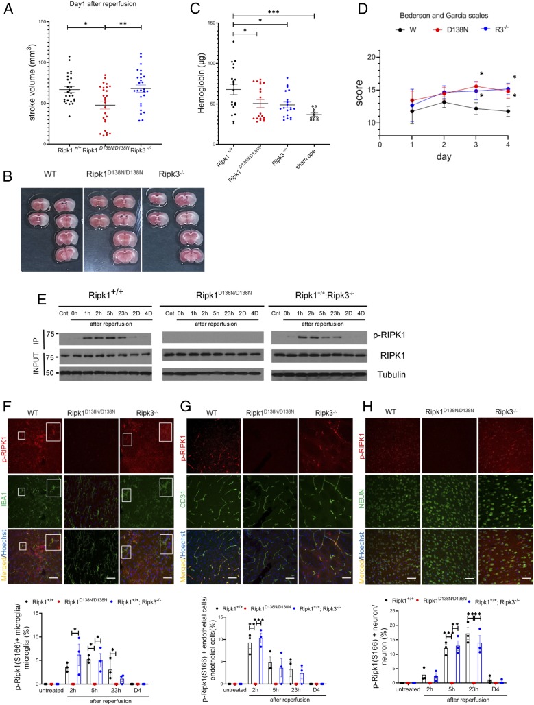
Apoptosis and necroptosis are two regulated cell death mechanisms; however, the interaction between these cell death pathways in vivo is unclear. Here we used cerebral ischemia/reperfusion as a model to investigate the interaction between apoptosis and necroptosis. We show that the activation of RIPK1 sequentially promotes necroptosis followed by apoptosis in a temporally specific manner. Cerebral ischemia/reperfusion insult rapidly activates necroptosis to promote cerebral hemorrhage and neuroinflammation. Ripk3 deficiency reduces cerebral hemorrhage and delays the onset of neural damage mediated by inflammation. Reduced cerebral perfusion resulting from arterial occlusion promotes the degradation of TAK1, a suppressor of RIPK1, and the transition from necroptosis to apoptosis. Conditional knockout of TAK1 in microglial/infiltrated macrophages and neuronal lineages sensitizes to ischemic infarction by promoting apoptosis. Taken together, our results demonstrate the critical role of necroptosis in mediating neurovascular damage and hypoperfusion-induced TAK1 loss, which subsequently promotes apoptosis and cerebral pathology in stroke and neurodegeneration.

Keywords: RIPK1; RIPK3; apoptosis; necroptosis; stroke.


Conflict of interest statement

Competing interest statement: J.Y. is a consultant of Denali Therapeutics Inc., which has licensed the necrostatin technology. M.P. received consulting and speaker fees from Genentech, GlaxoSmithKline, Boehringer Ingelheim, and Sanofi. All other authors declare no competing interests.

Figures

Fig. 1.
Fig. 1.
Activation of RIPK1 by ischemic insult. (A) WT, Ripk1D138N/D138N, and Ripk3−/− mice were subjected to transient 60-min MCAO, followed by reperfusion. The mice were killed after 23 h of reperfusion, and the brains were processed for TTC staining to measure infarct volume (n = 27 per group). (B) Representative TTC stainings from mice subjected to 60 min of ischemia followed by 23 h of reperfusion. (C) Quantification of hemorrhagic lesions by hemoglobin quantification in WT, Ripk1D138N/D138N, and Ripk3−/− mice treated as in A. (D) WT, Ripk1D138N/D138N, and Ripk3−/− mice were subjected to transient 60 min of MCAO followed by reperfusion, and were evaluated with Bederson and Garcia scales in a double-blinded manner daily from day 1 to day 4 after the MCAO procedure (n = 12 per group). The highest score is 21 points, and a higher score is correlated with better outcome. (E) Brain lysates from mice with indicated genotypes subjected to MCAO for 60 min followed by reperfusion for the indicated time periods were immunoprecipitated with p-S166 RIPK1 and then analyzed by immunoblotting with the indicated antibodies. (FH, Upper) Sections of WT, Ripk1D138N/D138N, and Ripk3−/− mice treated with transient 60 min of MCAO followed by reperfusion for 23 h (F and H) or 3 h (G) and at different time points as indicated on the bar graphs were immunostained with p-S166 RIPK1 and coimmunostained with IBA1, CD31, or NeuN for IBA1+ microglia/infiltrated macrophages (F), cerebrovascular endothelial cells (G), and neurons (H), respectively. (Magnification: 40×; Sale bar: 50 μm.) (FH, Lower) The quantification of the results. The data represent mean ± SEM. n = 3 mice for each time point. Assessment of each time point includes data from five sections for approximately 150 cells in each mouse. (F) The large square Insets (Right) are enlarged images of the small square Insets (Left). *P < 0.05, **P < 0.01, and ***P < 0.001.
Fig. 2.
Fig. 2.
Activation of necroptosis and apoptosis by ischemic insult. (A) Brain lysates from WT mice treated with MCAO for 60 min, followed by reperfusion for the indicated time periods were analyzed by immunoblotting for p-S345 MLKL, MLKL, p-T231/S232 RIPK3, RIPK3, CC3, and Tubulin. (B) Brain lysates from WT mice treated with permanent occlusion for the indicated time periods were analyzed by immunoblotting for p-S345 MLKL, MLKL, and Tubulin. (C) Brain lysates prepared in 6 M urea from WT mice treated with MCAO for 60 min followed by reperfusion for 0, 1, 2, or 5 h or without reperfusion were analyzed by immunoblotting for p-S345 MLKL and MLKL. (DE, Upper) Brain sections of WT, Ripk1D138N/D138N, and Ripk3−/− mice treated with MCAO for 60 min followed by reperfusion for 3 h were immunostained for p-T231/S232 RIPK3 (D) and p-S345 MLKL (E) and coimmunostained with CD31. P-RIPK3+ endothelial cells at different time points were quantified (Bottom). (FH) Brain sections of WT, Ripk1D138N/D138N, and Ripk3−/− mice treated with MCAO for 60 min followed by reperfusion for 23 h were immunostained for p-T231/S232 RIPK3 (F), p-S345 MLKL (G), or CC3 (H) and coimmunostained with NeuN. (DH, Lower) P-RIPK3+, p-MLKL+, and CC3+ neurons or endothelial cells as indicated at different time points were quantified. The data represent mean ± SEM; n= 3 mice per time point. (Magnification: 40×; Scale bar: 50 μm.) Assessment of each time point includes data from five sections for approximately 150 cells from each mouse. *P < 0.05, **P < 0.01, and ***P < 0.001.
Fig. 3.
Fig. 3.
Inhibition of RIPK1 blocks neuroinflammation induced by cerebral ischemic insult. Quantitative RT-PCR analysis of the mRNA expression levels of indicated cytokines and chemokines in the brain lysates from WT, Ripk1D138N/D138N, and Ripk3−/− mice treated with MCAO for 60 min followed by reperfusion for the indicated time periods (n = 5 mice for each time point). The data represent mean ± SEM. *P < 0.05.
Fig. 4.
Fig. 4.
Reduction of TAK1 in neurons and microglia/infiltrated macrophages induced by cerebral hypoperfusion. (A) Brain lysates from WT mice treated with MCAO for 60 min followed by reperfusion for the indicated time periods were analyzed by immunoblotting for TAK1, p-(T180/Y182) p38, p38, p-T183/Y185 JNK, JNK, p-T202/Y204 ERK, and ERK, with Tubulin as a loading control. (B) Brain lysates from mice with indicated genotypes subjected to MCAO for 60 min followed by reperfusion for the indicated time periods were analyzed by immunoblotting for TAK1 and Tubulin as a loading control. The same WT Ripk1+/+ data in A were used in B to compare with that of mutant mice. (C and D, Upper) Brain sections from WT mice subjected to MCAO for 60 min followed by reperfusion for 23 h were immunostained with TAK1 and coimmunostained with IBA1 (C) and NeuN (D). The nuclei were stained by Hoechst stain. Quantification of TAK1+/IBA1+ microglia/infiltrated macrophages and TAK1+/NeuN+ neurons are shown at the bottom of each panel. n = 3 mice. Quantifications include about 100 TAK1+ cells and about 100 CC3+ cells from each mouse. The fourth panels in each column are enlarged images of the Insets. (Scale bar, white: 100 μm; Magnification: 20×.) (Scale bar, yellow: 50 μm; Magnification: 40×.) (E, Left) TAK1+/CC3+ cells at 24 h after MCAO were quantified by double immunostaining for TAK1 and CC3. n = 3 mice. (E, Right) Quantifications included ∼100 cells. The data represent mean ± SEM. The fourth panels in each column are enlarged images of the Insets. (Scale bar, white: 100 μm; Magnification: 20×.) (Scale bar, yellow: 50 μm; Magnification: 40×.) (FI) Primary microglia (F and H) and neurons (G and I) from WT mice were treated with serum deprivation in the presence of MG132 (10 μM), E64d (5 μg/mL), Velcade (50 ng/mL), chloroquine (50 μg/mL), or a combination as indicated for 18 h. The lysates were analyzed by immunoblotting for TAK1, RIPK1, and Tubulin. (J) TTC staining of the mice with >80% rCBF reduction (Left) and <80% rCBF reduction (Right). Brain lysates from WT mice treated with no operation, sham operation, MCAO with >80% rCBF reduction, and MCAO with <80% rCBF reduction followed by reperfusion for 23 h were analyzed by immunoblotting for TAK1 and Tubulin as a loading control. *P < 0.05, **P < 0.01, and ***P < 0.001.
Fig. 5.
Fig. 5.
Selective TAK1 loss augments the activation of RIPK1 to promote apoptosis. (A) WT, MAP3K7fl/fl; Lyz2cre/+, and MAP3K7fl/fl; Emxcre/+ mice (each dot representing 1 mouse) were subjected to transient 60 min of MCAO, followed by reperfusion for 23 h. The brains were processed for TTC staining to measure infarct volume. (B) Brain lysates from mice with the indicated genotypes subjected to MCAO for 60 min followed by reperfusion for the indicated time periods were immunoprecipitated with p-S166 RIPK1 and analyzed by immunoblotting with the indicated antibody. (CE, Upper) Sections of WT, MAP3K7fl/fl; Lyz2cre/+, and MAP3K7fl/fl; Emxcre/+ mice treated with transient 60 min of MCAO followed by reperfusion for 5 h (C and E) or 3 h (D) at different time points as indicated on the bar graphs were immunostained with p-S166 RIPK1 and coimmunostained with IBA1, CD31, or NeuN for microglia/infiltrated macrophages (C), cerebrovascular endothelial cells (D), and neurons (E), respectively. The larger Insets (Right) in C are an enlarged image of the smaller Insets (Left). (Magnification: 40×; Scale bar: 50 μm.) (CE, Lower) The quantification of the results. The data represent mean ± SEM. The quantifications of p-S166 Ripk1+ microglia/infiltrated macrophages, endothelial cells, and neurons are shown at the bottom of each panel. n = 3 mice for each time point. Assessment of each time point includes data from five sections for approximately 150 cells in each mouse. *P < 0.05 and **P < 0.01.
Fig. 6.
Fig. 6.
Selective loss of TAK1 from microglia/infiltrated macrophages or neurons promotes apoptosis. (A) The brain lysates from mice with indicated genotypes reperfused for indicated periods after 60 min of MCAO were analyzed by immunoblotting for CC3 and Tubulin. (B, Upper) Brain sections of WT, MAP3K7fl/fl; Lyz2cre/+, and MAP3K7fl/fl; Emxcre/+ mice treated with MCAO for 60 min followed by reperfusion for 23 h were immunostained for CC3. (B, Lower) Quantifications of CC3+ neurons is shown at the bottom. n = 3 mice for each time point. The data represent mean ± SEM. Assessment of each time point includes data from five sections for approximately 150 cells in each mouse. (Magnification: 40×; Scale bar: 50 μm.) (C) Quantitative RT-PCR analysis of the mRNA expression of indicated cytokines and chemokines in the brains of WT, MAP3K7fl/fl; Lyz2cre/+, and MAP3K7fl/fl; Emxcre/+ mice subjected to 60 min of MCAO followed by reperfusion at different time periods. n = 4 mice per time point. The data represent mean ± SEM. (D) MAP3K7fl/fl; Lyz2cre/+ mice were subjected to 60 min of MCAO and then given vehicle only or Nec-1s(20 mg/kg) i.p. at 0 h and 17 h after the onset of MCAO. The brain lysates from MAP3K7fl/fl; Lyz2cre/+ mice reperfused for 23 h after 60 min occlusion were analyzed by immunoblotting for CC3 and Tubulin. (E) MAP3K7fl/fl; Emxcre/+ mice were subjected to 60 min of MCAO and then given vehicle only or Nec-1s(20 mg/kg) i.p. at 0 h and 17 h after the onset of MCAO. The brain lysates from MAP3K7fl/fl; Emxcre/+ mice reperfused for 23 h after 60 min of occlusion were analyzed by immunoblotting for CC3 and Tubulin. *P < 0.05, **P < 0.01, and ***P < 0.001.

References

    1. Moskowitz M. A., Lo E. H., Iadecola C., The science of stroke: Mechanisms in search of treatments. Neuron 67, 181–198 (2010). - PMC - PubMed
    1. Degterev A., et al. , Chemical inhibitor of nonapoptotic cell death with therapeutic potential for ischemic brain injury. Nat. Chem. Biol. 1, 112–119 (2005). - PubMed
    1. Yuan J., Yankner B. A., Apoptosis in the nervous system. Nature 407, 802–809 (2000). - PubMed
    1. Degterev A., et al. , Identification of RIP1 kinase as a specific cellular target of necrostatins. Nat. Chem. Biol. 4, 313–321 (2008). - PMC - PubMed
    1. Degterev A., Ofengeim D., Yuan J., Targeting RIPK1 for the treatment of human diseases. Proc. Natl. Acad. Sci. U.S.A. 116, 9714–9722 (2019). - PMC - PubMed

Publication types

Substances