. 2020 Mar 3;117(9):4724-4731.
doi: 10.1073/pnas.1917726117. Epub 2020 Feb 18.

Phage liquid crystalline droplets form occlusive sheaths that encapsulate and protect infectious rod-shaped bacteria

Affiliations

Phage liquid crystalline droplets form occlusive sheaths that encapsulate and protect infectious rod-shaped bacteria

Abul K Tarafder et al. Proc Natl Acad Sci U S A. .

Abstract

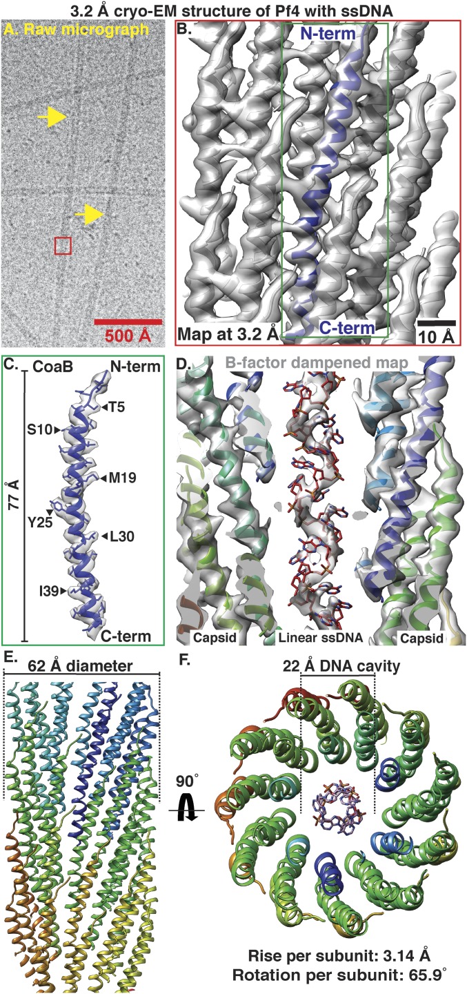
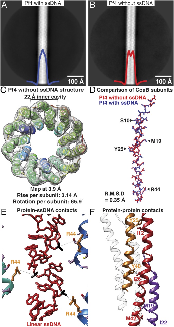
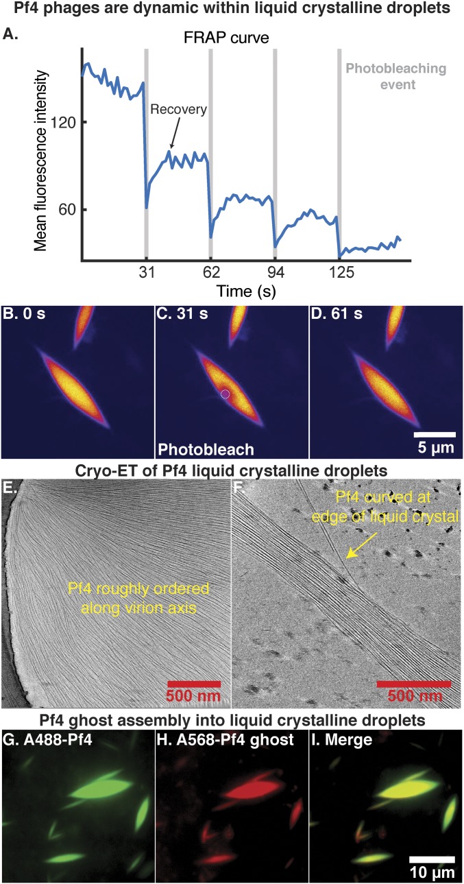
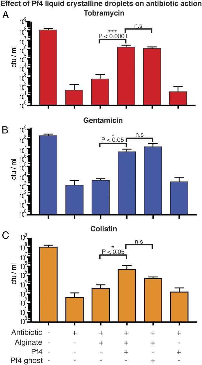
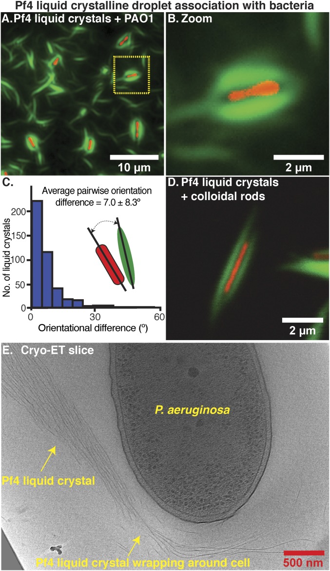
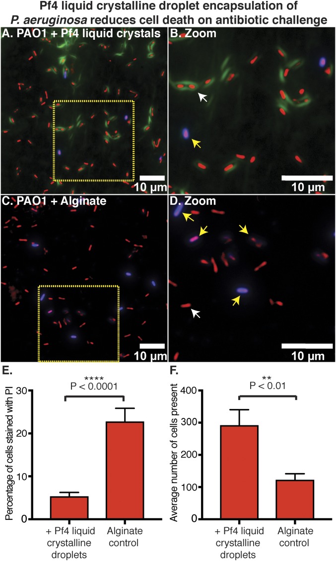
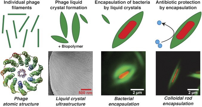
The opportunistic pathogen Pseudomonas aeruginosa is a major cause of antibiotic-tolerant infections in humans. P. aeruginosa evades antibiotics in bacterial biofilms by up-regulating expression of a symbiotic filamentous inoviral prophage, Pf4. We investigated the mechanism of phage-mediated antibiotic tolerance using biochemical reconstitution combined with structural biology and high-resolution cellular imaging. We resolved electron cryomicroscopy atomic structures of Pf4 with and without its linear single-stranded DNA genome, and studied Pf4 assembly into liquid crystalline droplets using optical microscopy and electron cryotomography. By biochemically replicating conditions necessary for antibiotic protection, we found that phage liquid crystalline droplets form phase-separated occlusive compartments around rod-shaped bacteria leading to increased bacterial survival. Encapsulation by these compartments was observed even when inanimate colloidal rods were used to mimic rod-shaped bacteria, suggesting that shape and size complementarity profoundly influences the process. Filamentous inoviruses are pervasive across prokaryotes, and in particular, several Gram-negative bacterial pathogens including Neisseria meningitidis, Vibrio cholerae, and Salmonella enterica harbor these prophages. We propose that biophysical occlusion mediated by secreted filamentous molecules such as Pf4 may be a general strategy of bacterial survival in harsh environments.

Keywords: Pseudomonas aeruginosa; antibiotic tolerance; cryo-EM; phage; phase separation.


Conflict of interest statement

The authors declare no competing interest.

Figures

Fig. 1.
Fig. 1.
Cryo-EM structure of Pf4 phage at 3.2-Å resolution. (A) Cryo-EM image of native Pf4 phage (yellow arrows) purified from biofilms (red box represents reconstructed segment). (B) Single-particle cryo-EM reconstruction of Pf4 phage (Movie S1). Cryo-EM density is shown as a gray isosurface, with the refined atomic coordinates of Pf4 CoaB protein subunits shown as ribbons (N- and C terminus of one CoaB marked). (C) Density and atomic model for a single CoaB protein. (D) Cross-section through the cryo-EM structure shows that the Pf4 ssDNA genome is linear. Map is B-factor dampened (50 Å2 compared to B and C) to aid visualization of the ssDNA. (E) Side view of Pf4 showing interdigitated arrangement of CoaB subunits within the capsid coat (only the front CoaB subunits displayed). (F) Top view of Pf4 showing linear ssDNA within the 22-Å inner cavity.
Fig. 2.
Fig. 2.
Cryo-EM structure of Pf4 filament without ssDNA at 3.9-Å resolution. (A) Two-dimensional class average of Pf4 with ssDNA, showing density in the core of the phage, indicated by peak in the horizontal density profile overlaid on average (blue curve). (B) Class average of Pf4 without ssDNA; a dip is observed in the horizontal density profile in the center of the average (red curve). (C) Top view of the cryo-EM structure of Pf4 without ssDNA at 3.9-Å resolution. Density is displayed as a gray isosurface and CoaB subunits as ribbons. The structure confirms lack of ssDNA in the 22-Å inner cavity. (D) Comparison of CoaB structure from Pf4 filaments with (blue) and without ssDNA (red) shows that the structures are almost identical (RMSD 0.35 Å, see Movie S2). (E) Magnified view of the CoaB:ssDNA interactions in the native Pf4 structure (from Fig. 1). Phosphates (black) of the linear ssDNA (red) are weakly coordinated by arginine 44 residues (orange) of CoaB (distance 5.4 Å). (F) Magnified view of CoaB:CoaB interactions within Pf4 filaments shows intersubunit hydrophobic interactions that stabilize the capsid.
Fig. 3.
Fig. 3.
Pf4 assembly into liquid crystalline droplets is dynamic with strong orientational ordering of filaments. (A) A region within a liquid crystalline droplet was photobleached multiple times and the FRAP was measured over time (Movie S3). Plot shows mean fluorescence intensity of the area targeted for photobleaching (y axis) against time (x axis). Photobleaching events are indicated by vertical gray bars. (BD) Fluorescent images at various time points during the FRAP experiment. Images have been background subtracted (yellow, high signal; blue, low signal). (Scale bar: BD, 5 μm.) (B) Pf4 liquid crystalline droplet before photobleaching. (C) Photobleaching of a region within the Pf4 liquid crystalline droplet indicated by the white circle. (D) Recovery of the fluorescence signal in the photobleached region indicating that Pf4 filaments are dynamic within the liquid crystal phase. (E and F) Cryo-ET of Pf4 liquid crystalline droplets (protein density black) shows longitudinal alignment of Pf4 filaments with the axis of the spindle (Movie S4). Curved Pf4 filaments are seen at the edges of the liquid crystalline droplet. (GI) Pf4 ghosts, with ssDNA chemically removed, can assemble into liquid crystalline droplets in the same manner as native Pf4. A488-Pf4 (green, G) and A568-Pf4 ghosts (red, H) were mixed with sodium alginate to form liquid crystalline droplets. Both compositional variants of Pf4 phage colocalized to the same liquid crystalline droplets (I). (Scale bar: GI, 10 μm.)
Fig. 4.
Fig. 4.
Pf4 liquid crystalline droplets with and without ssDNA protect P. aeruginosa cells against antibiotics. (A) Bar graph shows colony-forming units (cfu) per ml, a measure of P. aeruginosa culture cell viability after tobramycin treatment (y axis) in the presence of different reagents (x axis). Pf4 (native) and Pf4 ghost (without ssDNA) liquid crystalline droplets protect P. aeruginosa against tobramycin to a significantly greater extent than with sodium alginate alone (P < 0.0001). However, no significant difference is observed between treatments with Pf4 liquid crystalline droplets and Pf4 ghost liquid crystalline droplets. Values shown are the mean of six independent experiments (error bars show SD). (B) Graph shows cfu/mL (y axis) in the presence of different reagents (x axis). Pf4 liquid crystalline droplets had a significant protective effect over sodium alginate alone (P < 0.05) against gentamicin, but there was no significant difference between Pf4 liquid crystalline droplets and Pf4 ghost liquid crystalline droplets. (C) Pf4 liquid crystalline droplets had a significant protective effect over sodium alginate alone (P < 0.05) against colistin, but there was no significant difference between Pf4 liquid crystalline droplets and Pf4 ghost liquid crystalline droplets. Values shown are the mean of three independent experiments (error bars show SD). n.s, not significant.
Fig. 5.
Fig. 5.
Pf4 liquid crystalline droplets form protective sheaths around P. aeruginosa cells. (A) Optical microscopy of the condition from the antibiotic protection assay presented in Fig. 4, containing P. aeruginosa cells, alginate, and Pf4 (Fig. 4A, bar 4, no antibiotic). Transmitted light channel shows bacteria (red pseudocolor) and green fluorescent channel shows Pf4 (green). (B) Zoom of cell shows close association of liquid crystalline droplet around the cell. (C) Histogram of pairwise orientational differences between bacterial cells and associated liquid crystalline droplets from automated segmentation of fluorescence images (n = 417). (D) Colloidal rods (with a similar morphology to bacteria) mixed with Pf4 liquid crystalline droplets show the same encapsulation effect. (E) Cryo-ET slice showing a Pf4 liquid crystal phase associated with and wrapping around a bacterial cell (Movie S5).
Fig. 6.
Fig. 6.
Pf4 liquid crystalline droplet encapsulation of P. aeruginosa prevents cell death on antibiotic treatment. (A) Optical microscopy of the antibiotic protection assay presented in Fig. 4, containing P. aeruginosa cells, alginate, Pf4, and tobramycin (Fig. 4A, bar 4) stained with PI. Bacteria are shown in red (pseudocolor of transmitted light channel), Pf4 liquid crystalline droplets in green, and PI staining in blue. (B) Zoom of A shows Pf4 liquid crystalline droplet encapsulated bacteria are not stained with PI (live cells indicated by white arrows and dead cells indicated by yellow arrows). (C) Optical microscopy of the condition lacking Pf4 filaments from the antibiotic protection assay presented in Fig. 4, containing P. aeruginosa cells, alginate, and tobramycin (Fig. 4A, bar 3) stained with PI. (D) Zoom of C shows staining of cells with PI. All fluorescence images were background subtracted. (E) Bar chart showing quantitation of PI staining with the percentage of cells stained with PI (y axis) in the presence or absence of Pf4 liquid crystalline droplets (x axis). (F) Bar chart showing the average number of cells observed per image, in randomly collected fields of the sample (y axis) in the presence or absence of Pf4 liquid crystalline droplets (x axis). Mean values are shown and the error bars denote SE. Experiment was performed in triplicate (n = 30 images).
Fig. 7.
Fig. 7.
Schematic model of the mechanism of Pf4 phage-mediated antibiotic tolerance revealed in this study. Individual Pf4 phage filaments self-assemble into higher-order dynamic spindle-shaped liquid crystalline droplets termed tactoids in the presence of biopolymers. Pf4 liquid crystalline droplets encapsulate P. aeruginosa cells forming occlusive nanochambers that increase P. aeruginosa survival upon antibiotic treatment. This process is profoundly influenced by shape and size complementarity between the bacterial cells and liquid crystals as Pf4 liquid crystalline droplets can also encapsulate inanimate colloidal rods of comparable size and shape to P. aeruginosa bacteria.

Comment in

  • Phage liquid crystals protect Pseudomonas.
    Hofer U. Hofer U. Nat Rev Microbiol. 2020 May;18(5):264-265. doi: 10.1038/s41579-020-0347-6. Nat Rev Microbiol. 2020. PMID: 32099077 No abstract available.
  • Bacteria suit up with virus armor.
    Chang YW. Chang YW. Proc Natl Acad Sci U S A. 2020 Mar 24;117(12):6297-6299. doi: 10.1073/pnas.2001931117. Epub 2020 Mar 9. Proc Natl Acad Sci U S A. 2020. PMID: 32152106 Free PMC article. No abstract available.

References

    1. Costerton J. W., Stewart P. S., Greenberg E. P., Bacterial biofilms: A common cause of persistent infections. Science 284, 1318–1322 (1999). - PubMed
    1. Hall-Stoodley L., Stoodley P., Evolving concepts in biofilm infections. Cell. Microbiol. 11, 1034–1043 (2009). - PubMed
    1. Høiby N., Bjarnsholt T., Givskov M., Molin S., Ciofu O., Antibiotic resistance of bacterial biofilms. Int. J. Antimicrob. Agents 35, 322–332 (2010). - PubMed
    1. Whiteley M., et al. , Gene expression in Pseudomonas aeruginosa biofilms. Nature 413, 860–864 (2001). - PubMed
    1. Burgener E. B., et al. , Filamentous bacteriophages are associated with chronic Pseudomonas lung infections and antibiotic resistance in cystic fibrosis. Sci. Transl. Med. 11, eaau9748 (2019). - PMC - PubMed

Publication types

MeSH terms

LinkOut - more resources