. 2020 Feb 18;3(1):79.
doi: 10.1038/s42003-020-0792-9.

Beta amyloid aggregates induce sensitised TLR4 signalling causing long-term potentiation deficit and rat neuronal cell death

Affiliations

Beta amyloid aggregates induce sensitised TLR4 signalling causing long-term potentiation deficit and rat neuronal cell death

Craig Hughes et al. Commun Biol. .

Abstract

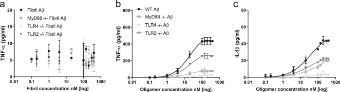
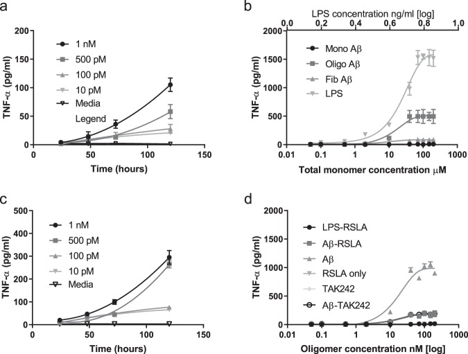
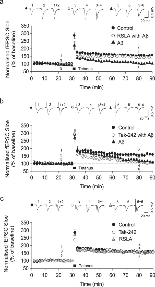
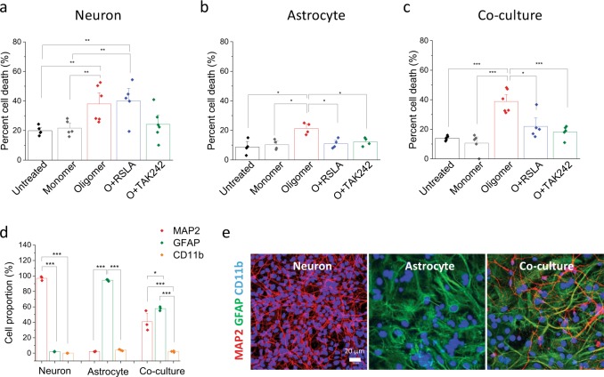
The molecular events causing memory loss and neuronal cell death in Alzheimer's disease (AD) over time are still unknown. Here we found that picomolar concentrations of soluble oligomers of synthetic beta amyloid (Aβ42) aggregates incubated with BV2 cells or rat astrocytes caused a sensitised response of Toll-like receptor 4 (TLR4) with time, leading to increased production of TNF-α. Aβ aggregates caused long term potentiation (LTP) deficit in hippocampal slices and predominantly neuronal cell death in co-cultures of astrocytes and neurons, which was blocked by TLR4 antagonists. Soluble Aβ aggregates cause LTP deficit and neuronal death via an autocrine/paracrine mechanism due to TLR4 signalling. These findings suggest that the TLR4-mediated inflammatory response may be a key pathophysiological process in AD.


Conflict of interest statement

The authors declare no competing interests.

Figures

Fig. 1
Fig. 1. Pro-inflammatory response of TLR4, MyD88 or TLR2 knockout macrophages to Abeta42(Aβ) oligomers and fibrils.
Cells (WT: wild type, MyD88−/−: MyD88 knockout, TLR4−/−: TLR4 knock out and TLR2−/−: TLR2 knockout) were stimulated with Aβ Fibrils (0.1–400 nM) and Aβ oligomers together with monomer (total monomer concentration 0.02–80 μM) for 24 h. The levels of the pro-inflammatory mediators TNF-α and IL-1β were measured. a TNF-α production remained unchanged at all concentrations with the addition of Fibrils (n = 5, ±sem). b TNF-α production significantly increases with increasing oligomer concentrations in all except the TLR4 knockout cells (n = 5, ±sem). c IL-1β levels production significantly increases with increasing oligomer concentrations in all except the TLR4 knockout cells (n = 5, mean ± sem).
Fig. 2
Fig. 2. Response of BV2 microglial cells and astrocytes to soluble Abeta42 (Aβ) aggregates (oligomers) and the blocking of the TNF-α production by TLR4 antagonists.
a Time course of TNF-α production by BV2 cells in response to continual exposure to soluble Aβ oligomers (10 pM–1 nM) together with monomer (monomer concentration 0.002–0.2 μM) (n = 4, sem). b The response of astrocytes to Aβ monomers, Aβ oligomers together with monomer (oligomer concentration 0.25 to 1000 nM and total monomer concentration 0.05–200 μM) and Aβ fibrils (total monomer concentration) compared to LPS (0.05–200 ng/ml) stimulation for 24 h (n = 4, mean ± sem). The astrocyte preparation had <2% microglial. c Time course of TNF-α production by astrocyte cells in response to continual exposure to Aβ oligomers (10 pM–1 nM) together with monomer (total monomer concentration 0.002–0.2 μM) (n = 4, mean ± sem). d The response of astrocytes after a 24 h incubation with Aβ oligomers (0.05–200 nM oligomer) together with monomer (total monomer 0.01–40 μM)) only and together with the TLR4 antagonist RSLA (0.1 µg/ml), LPS and RSLA, RSLA only and the TLR4 antagonist TAK-242 (1 µM) alone and with Aβ oligomers and monomer (n = 4, mean ± sem).
Fig. 3
Fig. 3. Aβ42-mediated inhibition of LTP is rescued by RSLA but not TAK-242 treatment.
Top: Example traces of fEPSPs from indicated time-points. Bottom: Mean fEPSP slope shown as percentage of the normalised baseline. a The inhibition of LTP by application of oligomerised Aβ42 (5 nM oligomers and 500 nM total monomer) was prevented by RSLA. b TAK-242 had no effect on Aβ-mediated inhibition of LTP. c TAK-242 and RSLA had no effect on the expression of LTP compared with control. All symbols represent the mean ± sem.
Fig. 4
Fig. 4. Neuronal cell death caused by Aβ42 oligomers is mediated by initiating TLR4 signalling by glial cells.
Cell death assay was performed using Sytox green on a live-cell imaging platform. a TLR4 inhibitors (0.1 µg/ml RSA, 1 µM TAK242) did not prevent Aβ42 oligomer-induced cell death in pure neuronal culture (5 nM oligomer and 1 µM total monomer). b, c Oligomer-induced astrocyte cell death was protected by TLR4 inhibitors. d, e Composition of the co-culture assessed using MAP2 (neuronal marker) and GFAP (astrocytic marker) immunocytochemistry together with representative images of a neuronal, astrocyte and co-culture. 97 ± 1.6% are MAP2-positive cells in neuronal prep, 92.2 ± 0.1% are GFAP-positive cells in astrocytic prep. 55.3 ± 7.1% MAP2 (+) and 44.1 ± 1.3% GFAP (+) are found in co-culture prep. The proportion of CD11b-positive cells is 3.8 ± 0.4% and 1.3 ± 0.5% in the astrocytic and co-culture preparations, respectively. (n = 5–6 from two independent experiments, mean ± sem).

References

    1. Shankar GM, et al. Amyloid-β protein dimers isolated directly from Alzheimerʼs brains impair synaptic plasticity and memory. Nat. Med. 2008;14:837. doi: 10.1038/nm1782. - DOI - PMC - PubMed
    1. Hong W, et al. Diffusible, highly bioactive oligomers represent a critical minority of soluble Aβ in Alzheimer’s disease brain. Acta Neuropathol. 2018;136:19–40. doi: 10.1007/s00401-018-1846-7. - DOI - PMC - PubMed
    1. Jo J, et al. Aβ1–42 inhibition of LTP is mediated by a signaling pathway involving caspase-3, Akt1 and GSK-3β. Nat. Neurosci. 2011;14:545. doi: 10.1038/nn.2785. - DOI - PubMed
    1. Li S, et al. Decoding the synaptic dysfunction of bioactive human AD brain soluble Aβ to inspire novel therapeutic avenues for Alzheimer’s disease. Acta Neuropathol. Commun. 2018;6:121. doi: 10.1186/s40478-018-0626-x. - DOI - PMC - PubMed
    1. Laurén J, Gimbel DA, Nygaard HB, Gilbert JW, Strittmatter SM. Cellular prion protein mediates impairment of synaptic plasticity by amyloid-β oligomers. Nature. 2009;457:1128. doi: 10.1038/nature07761. - DOI - PMC - PubMed

Publication types

MeSH terms