. 2020 Aug;46(5):478-492.
doi: 10.1111/nan.12610. Epub 2020 Apr 7.

Brain tumour diagnostics using a DNA methylation-based classifier as a diagnostic support tool

Affiliations

Brain tumour diagnostics using a DNA methylation-based classifier as a diagnostic support tool

L P Priesterbach-Ackley et al. Neuropathol Appl Neurobiol. 2020 Aug.

Abstract

Aims: Methylation profiling (MP) is increasingly incorporated in the diagnostic process of central nervous system (CNS) tumours at our centres in The Netherlands and Scandinavia. We aimed to identify the benefits and challenges of MP as a support tool for CNS tumour diagnostics.

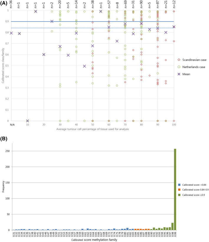
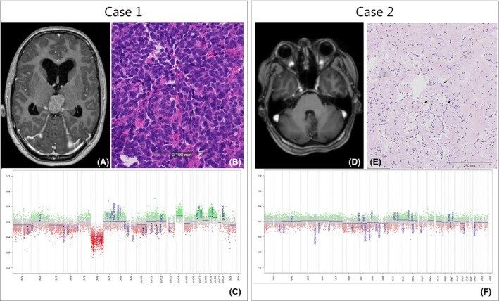
Methods: About 502 CNS tumour samples were analysed using (850 k) MP. Profiles were matched with the DKFZ/Heidelberg CNS Tumour Classifier. For each case, the final pathological diagnosis was compared to the diagnosis before MP.

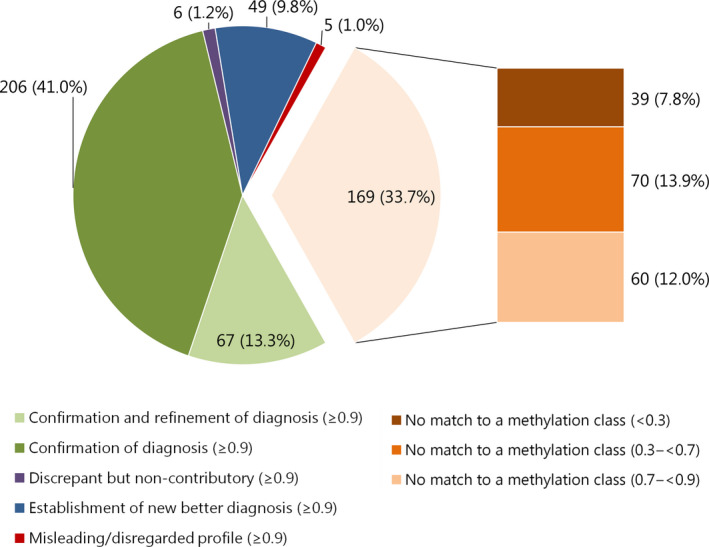
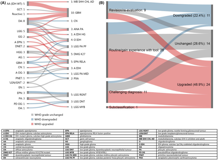
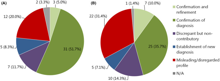
Results: In 54.4% (273/502) of all analysed cases, the suggested methylation class (calibrated score ≥0.9) corresponded with the initial pathological diagnosis. The diagnosis of 24.5% of these cases (67/273) was more refined after incorporation of the MP result. In 9.8% of cases (49/502), the MP result led to a new diagnosis, resulting in an altered WHO grade in 71.4% of these cases (35/49). In 1% of cases (5/502), the suggested class based on MP was initially disregarded/interpreted as misleading, but in retrospect, the MP result predicted the right diagnosis for three of these cases. In six cases, the suggested class was interpreted as 'discrepant but noncontributory'. The remaining 33.7% of cases (169/502) had a calibrated score <0.9, including 7.8% (39/502) for which no class indication was given at all (calibrated score <0.3).

Conclusions: MP is a powerful tool to confirm and fine-tune the pathological diagnosis of CNS tumours, and to avoid misdiagnoses. However, it is crucial to interpret the results in the context of clinical, radiological, histopathological and other molecular information.

Keywords: central nervous system tumours; diagnostics; methylation profiling.


Conflict of interest statement

The authors declare that they have no conflict of interest.

Figures

Figure 1
Figure 1
(A) Effect of estimated tumour cell percentage on calibrated score: Scatter plot of average tumour cell percentage (x‐axis) versus calibrated score for MP class family (Y‐axis). Cases from NL: green circles; cases from Scandinavia: red diamonds; purple crosses: mean. Horizontal lines: blue solid ‐ threshold of calibrated score ≥0.9; blue dashed ‐possible alternative threshold for calibrated score at ≥0.84 as suggested by [6] Cases with ‘no match <0.3’ (no calibrated score provided) were given the value 0 to be able to visualize them in this plot. The mean calibrated score of samples for which no tumour cell percentage was available is plotted at the bottom of the x‐axis, marked with ‘N/A’. NB. Symbols are often superimposed; labels at the top show the number of plotted cases. (B) Distribution of cases by calibrated scores for methylation class family: bar chart showing frequency of cases (Y‐axis) with specified calibrated score (X‐axis) for 468 cases with MP result. Valid matches with calibrated score ≥0.9 presented in green; no match cases with calibrated scores 0.84‐<0.9 in orange, remaining no match cases with calibrated scores 0.31‐<0.84 in blue. Data on no match cases with scores < 0.3 are not shown.
Figure 2
Figure 2
Effect of methylation profiling on diagnosis signed out to the clinicians for 502 cases with a calibrated score ≥0.9: light orange pie section represents all ‘no match’ cases combined. These are subdivided into calibrated scores <0.3; 0.3 to <0.7 and 0.7 to <0.9 in the bar to the right of this pie section. Labels represent: n (%) of 502 cases total.
Figure 3
Figure 3
New diagnoses after methylation profiling: (A) Overview of initial (left) and new diagnoses after MP (right) in cases classified as ‘Establishing new better diagnosis (≥0.9)’ (n = 49). (B) WHO grade effects in cases with establishment of new diagnosis: Difference between WHO grade original diagnosis and final diagnosis for cases categorized as ‘Establishment of new better diagnosis (≥0.9)’ (n = 49), subdivided by reasons to perform MP. Green shades: downgraded, blue shades: unchanged, red shades: upgraded.
Figure 4
Figure 4
(A) Effect of methylation profiling on final pathological diagnosis of 60 cases with calibrated score of 0.7 to <0.9 for the methylation class family: N/A refers to unresolved cases. Labels represent: n (%) of 60 cases total. (B) Effect of methylation profiling on final pathological diagnosis of 70 cases with calibrated score of 0.3 to <0.7 for the methylation class family: N/A refers to unresolved cases. Labels represent: n (%) of 70 cases total.
Figure 5
Figure 5
Case 1. (A) MR image, T1‐weighted after IV Gadolinium‐based contrast administration: tumour in the pineal region of a 15‐year‐old girl. (B) H&E stain 10×. (C) MP CNV plot, showing loss of chromosome 6. Case 2. (D) MR image, T1‐weighted after IV Gadolinium showing a tumour in cerebellum with close relation to the cerebral aqueduct and brainstem. (E) H&E stains 10×, arrows indicating focal perivascular pseudorosettes. (F) CNV plot showing a flat baseline with no indication of chromosomal changes in this tumour. Scale bars indicate (B) 100 μm; (E) 250 μm.

References

    1. Kros JM, Gorlia T, Kouwenhoven MC, Zheng P, Collins VP, Figarella‐branger D et al Panel review of anaplastic oligodendroglioma from European organization for research and treatment of cancer trial 26951: assessment of consensus in diagnosis, influence of 1p/19q Loss, and correlations with outcome. J Neuropathol Exp Neurol 2007; 66: 545–51 - PubMed
    1. Kristensen BW, Priesterbach‐Ackley LP, Petersen JK, Wesseling P. Molecular pathology of tumors of the central nervous system. Ann Oncol 2019; 30: 1265–78 - PMC - PubMed
    1. Noushmehr H, Weisenberger DJ, Diefes K, Phillips HS, Pujara K, Berman BP et al Identification of a CpG Island methylator phenotype that defines a distinct subgroup of glioma. Cancer Cell 2010; 17: 510–22 - PMC - PubMed
    1. Danielsson A, Nemes S, Tisell M, Lannering B, Nordborg C, Sabel M et al MethPed: a DNA methylation classifier tool for the identification of pediatric brain tumor subtypes. Clin Epigenetics 2015; 7: 62 - PMC - PubMed
    1. Capper D, Jones DTW, Sill M, Hovestadt V, Schrimpf D, Sturm D et al DNA‐methylation‐based classification of central nervous system tumours. Nature. 2018; 555: 469–474. 10.1038/nature26000 - DOI - PMC - PubMed