

Comparative Study
doi: 10.1148/radiol.2020200432.
Epub 2020 Feb 19.
Sensitivity of Chest CT for COVID-19: Comparison to RT-PCR
Affiliations
- PMID: 32073353
- PMCID: PMC7233365
- DOI: 10.1148/radiol.2020200432
Item in Clipboard
Comparative Study
Sensitivity of Chest CT for COVID-19: Comparison to RT-PCR
Radiology.
2020 Aug.
No abstract available
Figures

Flowchart for patient inclusion.

Examples of typical chest CT findings compatible with COVID-19 pneumonia in patients with epidemiological and clinical presentation suspicious for COVID-19 infection. A, male, 74 years old with fever and cough for 5 days. Axial chest CT shows bilateral subpleural ground glass opacities (GGO). B, female, 55 years old, with fever and cough for 7 days. Axial chest CT shows extensive bilateral ground glass opacities and consolidation; C, male, 43 years old, presenting with fever and cough for 1 week. Axial chest CT shows small bilateral areas of peripheral GGO with minimal consolidation; D, female, 43 years old presenting with fever with cough for 5 days. Axial chest CT shows a right lung region of peripheral consolidation.

Examples of typical chest CT findings compatible with COVID-19 pneumonia in patients with epidemiological and clinical presentation suspicious for COVID-19 infection. A, male, 74 years old with fever and cough for 5 days. Axial chest CT shows bilateral subpleural ground glass opacities (GGO). B, female, 55 years old, with fever and cough for 7 days. Axial chest CT shows extensive bilateral ground glass opacities and consolidation; C, male, 43 years old, presenting with fever and cough for 1 week. Axial chest CT shows small bilateral areas of peripheral GGO with minimal consolidation; D, female, 43 years old presenting with fever with cough for 5 days. Axial chest CT shows a right lung region of peripheral consolidation.
Examples of typical chest CT findings compatible with COVID-19 pneumonia in patients with epidemiological and clinical presentation suspicious for COVID-19 infection. A, male, 74 years old with fever and cough for 5 days. Axial chest CT shows bilateral subpleural ground glass opacities (GGO). B, female, 55 years old, with fever and cough for 7 days. Axial chest CT shows extensive bilateral ground glass opacities and consolidation; C, male, 43 years old, presenting with fever and cough for 1 week. Axial chest CT shows small bilateral areas of peripheral GGO with minimal consolidation; D, female, 43 years old presenting with fever with cough for 5 days. Axial chest CT shows a right lung region of peripheral consolidation.

Examples of typical chest CT findings compatible with COVID-19 pneumonia in patients with epidemiological and clinical presentation suspicious for COVID-19 infection. A, male, 74 years old with fever and cough for 5 days. Axial chest CT shows bilateral subpleural ground glass opacities (GGO). B, female, 55 years old, with fever and cough for 7 days. Axial chest CT shows extensive bilateral ground glass opacities and consolidation; C, male, 43 years old, presenting with fever and cough for 1 week. Axial chest CT shows small bilateral areas of peripheral GGO with minimal consolidation; D, female, 43 years old presenting with fever with cough for 5 days. Axial chest CT shows a right lung region of peripheral consolidation.

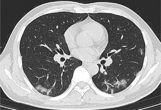
Examples of chest CT findings less commonly reported in COVID-19 infection (atypical) in patients with epidemiological and clinical presentation suspicious for COVID-19 infection. A, male, 36 years old with cough for 3 days. Axial chest CT shows a small focal and central ground glass opacity (GGO) in the right upper lobe; B, female, 40 years old. Axial chest CT shows small peripheral linear opacities bilaterally. C, male, 38 years old. Axial chest CT shows a GGO in the central left lower lobe; D, male, 31 years old with fever for 1 day. Axial chest CT shows a linear opacity in the left lower lateral mid lung.

Examples of chest CT findings less commonly reported in COVID-19 infection (atypical) in patients with epidemiological and clinical presentation suspicious for COVID-19 infection. A, male, 36 years old with cough for 3 days. Axial chest CT shows a small focal and central ground glass opacity (GGO) in the right upper lobe; B, female, 40 years old. Axial chest CT shows small peripheral linear opacities bilaterally. C, male, 38 years old. Axial chest CT shows a GGO in the central left lower lobe; D, male, 31 years old with fever for 1 day. Axial chest CT shows a linear opacity in the left lower lateral mid lung.

Examples of chest CT findings less commonly reported in COVID-19 infection (atypical) in patients with epidemiological and clinical presentation suspicious for COVID-19 infection. A, male, 36 years old with cough for 3 days. Axial chest CT shows a small focal and central ground glass opacity (GGO) in the right upper lobe; B, female, 40 years old. Axial chest CT shows small peripheral linear opacities bilaterally. C, male, 38 years old. Axial chest CT shows a GGO in the central left lower lobe; D, male, 31 years old with fever for 1 day. Axial chest CT shows a linear opacity in the left lower lateral mid lung.

Examples of chest CT findings less commonly reported in COVID-19 infection (atypical) in patients with epidemiological and clinical presentation suspicious for COVID-19 infection. A, male, 36 years old with cough for 3 days. Axial chest CT shows a small focal and central ground glass opacity (GGO) in the right upper lobe; B, female, 40 years old. Axial chest CT shows small peripheral linear opacities bilaterally. C, male, 38 years old. Axial chest CT shows a GGO in the central left lower lobe; D, male, 31 years old with fever for 1 day. Axial chest CT shows a linear opacity in the left lower lateral mid lung.
Comment in
-
Recommendation of low-dose CT in the detection and management of COVID-2019.Eur Radiol. 2020 Aug;30(8):4356-4357. doi: 10.1007/s00330-020-06809-6. Epub 2020 Mar 19. Eur Radiol. 2020. PMID: 32193637 Free PMC article. No abstract available.
-
A British Society of Thoracic Imaging statement: considerations in designing local imaging diagnostic algorithms for the COVID-19 pandemic.Clin Radiol. 2020 May;75(5):329-334. doi: 10.1016/j.crad.2020.03.008. Clin Radiol. 2020. PMID: 32265036 Free PMC article. No abstract available.
References
-
- Song F, Shi N, Shan F, et al. Emerging Coronavirus 2019-nCoV Pneumonia. Radiology 2020. doi:10.1148/radiol.2020200274. Published online February 6, 2020.
Publication types
MeSH terms
LinkOut - more resources
Full Text Sources
Other Literature Sources
Medical
