. 2020 May;35(4):284-291.
doi: 10.1089/cbr.2019.3079. Epub 2020 Feb 19.

Comparison of Prostate-Specific Membrane Antigen Expression Levels in Human Salivary Glands to Non-Human Primates and Rodents

Affiliations

Comparison of Prostate-Specific Membrane Antigen Expression Levels in Human Salivary Glands to Non-Human Primates and Rodents

Jyoti Roy et al. Cancer Biother Radiopharm. 2020 May.

Abstract

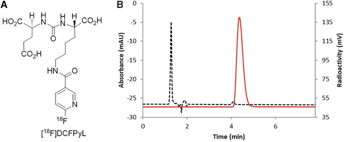
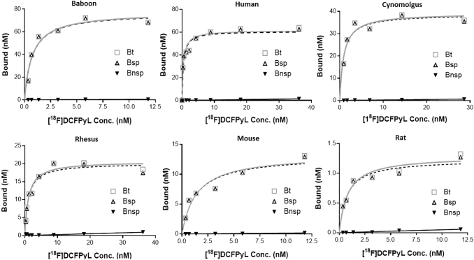
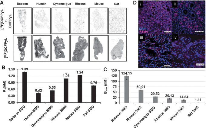
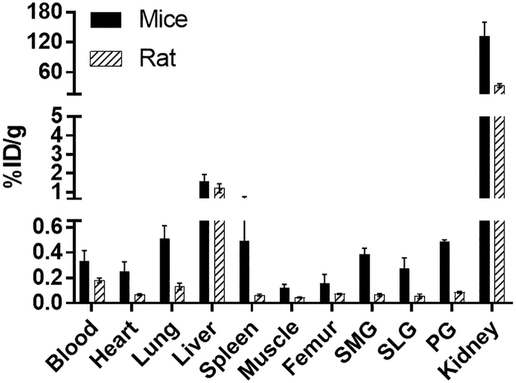
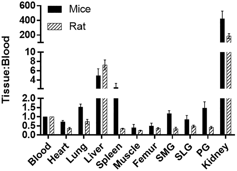
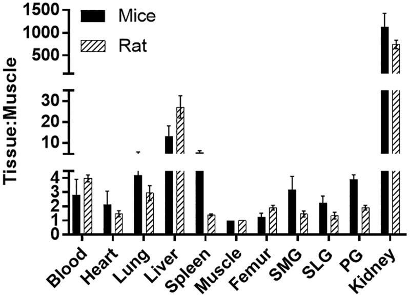
Background: Prostate-specific membrane antigen (PSMA) has emerged as a promising target for developing radionuclide therapy (RNT) in prostate cancer; however, accumulation of PSMA-RNT in salivary glands can result in irreversible xerostomia. Methods to prevent PSMA-RNT-related xerostomia could be clinically useful; however, little is known about PSMA expression in salivary glands of preclinical animal models. Using [18F]DCFPyL autoradiography/biodistribution, PSMA expression levels were determined in salivary glands of various preclinical monkey and rodent species and compared with humans. Methods: Binding affinities (Kd) and PSMA levels (Bmax) were determined by in vitro [18F]DCFPyL autoradiography studies. In vivo rodent tissue uptakes (%ID/g) were determined from [18F]DCFPyL biodistributions. Results: [18F]DCFPyL exhibited low nanomolar Kd for submandibular gland (SMG) PSMA across all the species. PSMA levels in human SMG (Bmax = 60.91 nM) were approximately two-fold lower compared with baboon SMG but were two- to three-fold higher than SMG PSMA levels of cynomolgus and rhesus. Rodents had the lowest SMG PSMA levels, with the mouse being 10-fold higher than the rat. In vivo rodent biodistribution studies confirmed these results. Conclusions: SMG of monkeys exhibited comparable PSMA expression to human SMG whereas rodents were lower. However, the results suggest that mice are relatively a better small animal preclinical model than rats for PSMA salivary gland studies.

Keywords: PET imaging; PSMA; autoradiography; radionuclide imaging; radionuclide therapy; salivary glands.


Conflict of interest statement

There are no potential conflicts of interest to disclose.

Figures

FIG. 1.
FIG. 1.
(A). Structure of [F]DCFPyL. (B). HPLC analysis of [F]DcFPyL (HPLC conditions: Agilent Eclipse plus C18 column (4.6 × 150 mm, 3.5 μm), mobile phase: 10% acetonitrile in 0.1 M ammonium formate, with a flow rate of 1.0 mL/min. Solid line: in-line radio detector; dotted line: UV detector at 254 nm.
FIG. 2.
FIG. 2.
Representative plots of in vitro autoradiography saturation binding studies of human, non-human primates (baboon, cynomolgus, rhesus) and rodent (mouse, rat) submandibular glands; each point (n = 6 points) is derived from ROI analysis. For each plot: Bt = Bound total; Bnsp = Bound non-specific; Bsp = Bound specific (Bt-Bnsp = Bsp). ROI, regions of interest.
FIG. 3.
FIG. 3.
(A) Representative autoradiograms showing [F]DCFPyL binding (Bt:[F]DCFPyL) and non-specific binding (Bnsp: [F]DCFPyL+DCFPyL (10−6M)) to PSMA in the SMGs of various species. (B, C) Binding affinity (Kd, nM) of [F]DCFPyL (B) and PSMA concentrations (C; Bmax, nM) in submandibular glands of various species determined from in vitro autoradiograph saturation plots. Each bar represents mean ± SD, n = 3; (D) Representative immunofluorescence staining of PC3-PSMA tumor (i), human submandibular glands (ii), baboon submandibular glands (iii), and mouse submandibular glands (iv) with PSMA antibody (PSMA expression, pink) and DAPI (nuclear stain, blue). SMG, submandibular gland; PSMA, prostate-specific membrane antigen.
FIG. 4.
FIG. 4.
Comparison of [F]DCFPyL biodistribution in rats and mice at 1 h after injection of [F]DCFPyL. Each bar represents %ID/g ± SD (for mice: %ID/g normalized to 20 g mice; for rats: %ID/g normalized to 125 g rat; n = 5 for each group). SMG, SLG, and PG represent submandibular, sublingual, and parotid glands, respectively.
FIG. 5.
FIG. 5.
Comparison of Tissue:Blood ratios of [F]DCFPyL in rats and mice at 1 h after injection of [F]DCFPyL. Each bar represents Tissue:Blood ± SD; n = 5 for each group. SMG, SLG, and PG represent submandibular, sublingual, and parotid glands, respectively.
FIG. 6.
FIG. 6.
Comparison of Tissue:Muscle ratios of [F]DCFPyL in rats and mice at 1 h after injection of [F]DCFPyL. Each bar represents Tissue:Muscle ± SD; n = 5 for each group. SMG, SLG, and PG represent submandibular, sublingual, and parotid glands, respectively.

References

    1. Davis MI, Bennett MJ, Thomas LM, et al. . Crystal structure of prostate-specific membrane antigen, a tumor marker and peptidase. Proc Natl Acad Sci U S A 2005;102:5981. - PMC - PubMed
    1. Sweat SD, Pacelli A, Murphy GP, et al. . Prostate-specific membrane antigen expression is greatest in prostate adenocarcinoma and lymph node metastases. Urology 1998;52:637. - PubMed
    1. Bostwick DG, Pacelli A, Blute M, et al. . Prostate specific membrane antigen expression in prostatic intraepithelial neoplasia and adenocarcinoma - A study of 184 cases. Cancer-Am Cancer Soc 1998;82:2256 - PubMed
    1. Rahbar K, Afshar-Oromieh A, Jadvar H, et al. . PSMA theranostics: Current status and future directions. Mol Imaging 2018;17 - PMC - PubMed
    1. Chakravarty R, Siamof CM, Dash A, et al. . Targeted alpha-therapy of prostate cancer using radiolabeled PSMA inhibitors: A game changer in nuclear medicine. Am J Nucl Med Mol Imaging 2018;8:247. - PMC - PubMed

LinkOut - more resources