. 2020 Feb 14;10(2):13.
doi: 10.3390/bios10020013.

Detection of the Ovarian Cancer Biomarker Lysophosphatidic Acid in Serum

Affiliations

Detection of the Ovarian Cancer Biomarker Lysophosphatidic Acid in Serum

Brian De La Franier et al. Biosensors (Basel). .

Abstract

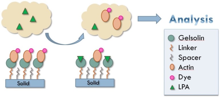
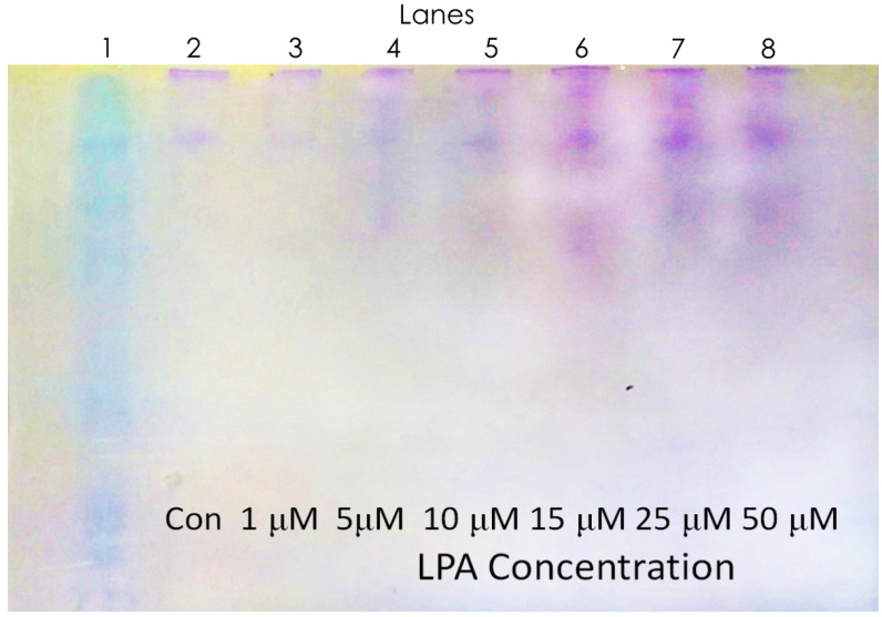
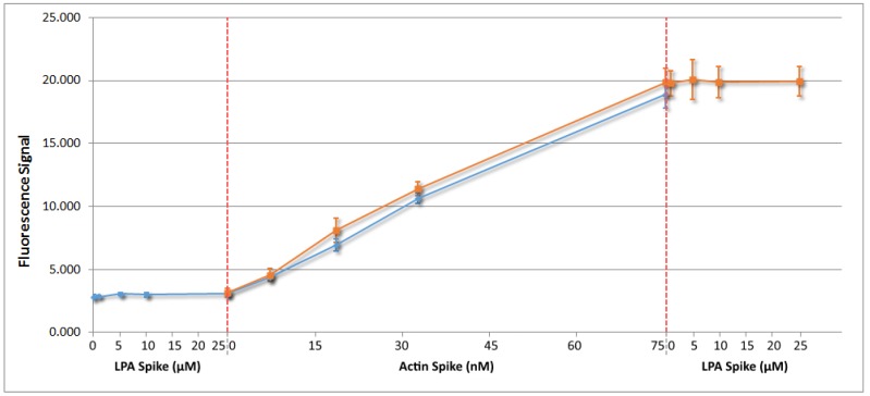
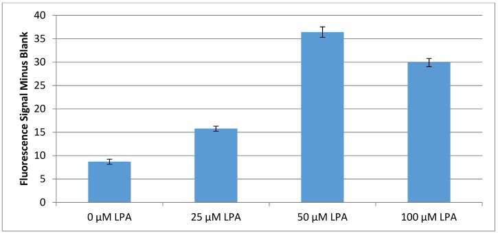
Lysophosphatidic acid (LPA) is present during the medical condition of ovarian cancer at all stages of the disease, and, therefore possesses considerable potential as a biomarker for screening its presence in female patients. Unfortunately, there is currently no clinically employable assay for this biomarker. In the present work, we introduce a test based on the duel protein system of actin and gelsolin that could allow the quantitative measurement of LPA in serum samples in a biosensing format. In order to evaluate this possibility, actin protein was dye-modified and complexed with gelsolin protein, followed by surface deposition onto silica nanoparticles. This solid-phase system was exposed to serum samples containing various concentrations of LPA and analyzed by fluorescence microscopy. Measurements conducted for the LPA-containing serum samples were higher after exposure to the developed test than samples without LPA. Early results suggest a limit of detection of 5 μM LPA in serum. The eventual goal is to employ the chemistry described here in a biosensor configuration for the large population-scale, rapid screening of women for the potential occurrence of ovarian cancer.

Keywords: actin; fluorescence detection; gelsolin; lysophosphatidic acid; ovarian cancer.


Conflict of interest statement

The authors declare no conflict of interest.

Figures

Figure 1
Figure 1
Principle behind actin–gelsolin chemistry for detection of LPA in patient samples. Solid support used in these experiments was silica gel.
Figure 2
Figure 2
Structures of surface bound 3-(3-(trichlorosilyl)propoxy)propanoyl chloride (MEG-Cl) (A) and perfluorophenyl 12-(trichlorosilyl)dodecanoate (PFP-TTTA) (B), both before and after extension with Ni-NTA (Nα,Nα-bis(carboxymethyl)-l-lysine with nickel).
Figure 3
Figure 3
A non-denaturing gel where mixtures of actin and gelsolin were run. Concentration ratios were chosen based on the dissolved concentration of actin.
Figure 4
Figure 4
A non-denaturing gel where each lane contains a 1:1 mixture of gelsolin and actin that has been incubated with a varying concentration of lysophosphatidic acid (LPA).
Figure 5
Figure 5
Fluorescence analysis of rhodamine-dyed actin in serum where in orange actin was first added to serum at the specified concentrations followed by the addition of LPA to the final serum solution, and in blue LPA was added to serum at the specified concentrations followed by the addition of actin to the final solution.
Figure 6
Figure 6
Fluorescence results for silica gel tests incubated in serum samples for 20 min.

Similar articles

Cited by

References

    1. De La Franier B., Thompson M. Biosensors and Methods for Detection of Lysophosphatidic Acid for Signaling of Ovarian Cancer. US 15/572,295. U.S. Pantent. 2018 May 31;
    1. Siegel R., Naishadham D., Jemal A. Cancer statistics for hispanics/latinos, 2012. CA Cancer J. Clin. 2012;62:283–298. doi: 10.3322/caac.21153. - DOI - PubMed
    1. Stevens G.A., Mathers C.D., Beard J.R. Global mortality trends and patterns in older women. Bull. World Health Organ. 2013;91:630–639. doi: 10.2471/BLT.12.109710. - DOI - PMC - PubMed
    1. Sant M., Allemani C., Santaquilani M., Knijn A., Marchesi F., Capocaccia R. EUROCARE-4. Survival of cancer patients diagnosed in 1995–1999. Results and commentary. Eur. J. Cancer. 2009;45:931–991. doi: 10.1016/j.ejca.2008.11.018. - DOI - PubMed
    1. Jemal A., Bray F., Center M.M., Ferlay J., Ward E., Forman D. Global cancer statistics. CA A Cancer J. Clin. 2011;61:69–90. doi: 10.3322/caac.20107. - DOI - PubMed