. 2020 Feb 15;8(1):19.
doi: 10.3390/dj8010019.

Management of a Facilitated Aesthetic Orthodontic Treatment with Clear Aligners and Minimally Invasive Corticotomy

Affiliations

Management of a Facilitated Aesthetic Orthodontic Treatment with Clear Aligners and Minimally Invasive Corticotomy

Silvia Caruso et al. Dent J (Basel). .

Abstract

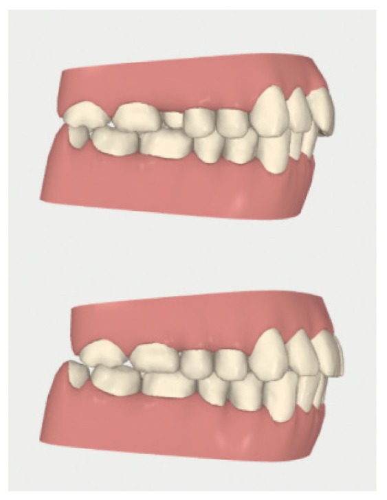
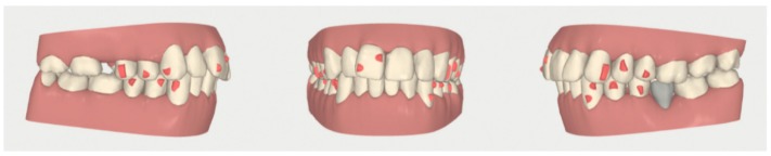
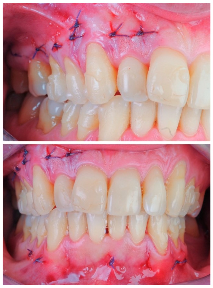
Accelerating orthodontic tooth movement has become a topical issue and the corticotomy seems to be the only effective and safe technique reported in the literature. Simultaneously, aesthetic orthodontic treatment with removable clear aligners has become commonly requested. The aim of this paper is to illustrate the management of facilitated aesthetic orthodontic treatment, a combined approach including piezocision corticotomy and clear aligners for orthodontic treatment. Orthodontic planning for traditional clear aligners should be modified to take advantage of the corticotomy technique in order to facilitate the most difficult orthodontic movements needed to achieve treatment completion, where each aligner will be used for four days rather than 15 days for a total time of four months. A corticotomy with a modified minimally invasive flapless piezocision technique should be performed in both jaws at the same time, before the time window of the orthodontic treatment, where the most difficult orthodontic movements are planned. Treatment planning where difficult orthodontic movements, such as anterior open-bite closure and extraction space closure, are easily managed with clear aligners and are presented as examples of facilitated aesthetic orthodontic treatment application. The combination between aesthetic treatment with clear aligners and modified piezocision corticotomy, if carefully planned, seems to represent a synergy that achieves the current goals of orthodontic treatment. The primary objectives of this combination should be facilitating difficult orthodontic movements and reducing treatment duration.

Keywords: aesthetics; clear aligner appliances; cortical bone injuries; orthodontic tooth movement; removable orthodontic appliances.


Conflict of interest statement

The authors declare no conflict of interest.

Figures

Figure 1
Figure 1
Cone Beam Computed Tomography (CBCT) image of modified piezocision corticotomy between 4-2 and 4-3.
Figure 2
Figure 2
Appropriate sonic insert to perform the modified piezocision technique (thickness: 0.25 mm).
Figure 3
Figure 3
Surgical procedure: (A) incision with a 15 blade; (B) cortical incision with sonic device and appropriate insert; (C) sutures with Vicryl 5.0 thread are applied only if bleeding persists.
Figure 4
Figure 4
Case 1: initial and final frontal and lateral photo.
Figure 5
Figure 5
Case 1: initial (A,B) and final (C,D) superior and inferior occlusal arches photo.
Figure 6
Figure 6
Case 1: cephalometric image.
Figure 7
Figure 7
Case 1: initial and final digital frontal and lateral images showing the adopted attachment placement.
Figure 8
Figure 8
Case 1: modified piezocision corticotomy detail.
Figure 9
Figure 9
Case 1: photos after the surgical procedure; sutures with Vicryl 5.0 thread were applied only where bleeding persisted.
Figure 10
Figure 10
Case 1: digital sequential (up to down) detail of the difficult radicular movement of 3-2 and 4-2.
Figure 11
Figure 11
Case 2: initial and final frontal and lateral photo.
Figure 12
Figure 12
Case 2: initial (A,B) and final (C,D) superior and inferior occlusal arches photos.
Figure 13
Figure 13
Case 2: orthopantomography.
Figure 14
Figure 14
Case 2: cephalometric image.
Figure 15
Figure 15
Case 2: initial and final digital occlusal images.
Figure 16
Figure 16
Case 2: digital sequential (left to right) detail of the facilitated space closure orthodontic movement.
Figure 17
Figure 17
Case 2: digital lateral views before and after the space closure.
Figure 18
Figure 18
Case 2: initial digital frontal and lateral images showing the appropriate attachment placement for achieving the space closure.
Figure 19
Figure 19
Case 2: photographs after the surgical procedure; sutures with Vicryl 5.0 thread were applied only where bleeding persisted.

References

    1. Cassetta M., Pandolfi S., Giansanti M. Minimally invasive corticotomy in orthodontics: A new technique using a CAD/CAM surgical template. Int. J. Oral Maxillofac. Surg. 2015;44:830–833. doi: 10.1016/j.ijom.2015.02.020. - DOI - PubMed
    1. Silvestrini Biavati A., Tecco S., Migliorati M., Festa F., Panza G., Marzo G., Gherlone E., Tetè S. Three-dimensional tomographic mapping related to primary stability and structural miniscrew characteristics. Orthod. Craniofac. Res. 2011;14:88–99. doi: 10.1111/j.1601-6343.2011.01512.x. - DOI - PubMed
    1. Liou E.J., Huang C.S. Rapid canine retraction through distraction of the periodontal ligament. Am. J. Orthod. Dentofac. Orthop. 1998;114:372–382. doi: 10.1016/S0889-5406(98)70181-7. - DOI - PubMed
    1. Collins M.K., Sinclair P.M. The local use of vitamin D to increase the rate of orthodontic tooth movement. Am. J. Orthod. Dentofac. Orthop. 1988;94:278–284. doi: 10.1016/0889-5406(88)90052-2. - DOI - PubMed
    1. Kalra A., Jaggi N., Bansal M., Goel S., Medsinge S.V., Abraham R., Jasoria G. Comparison of rate of canine retraction into recent extraction site with and without gingival fiberotomy: A clinical study. J. Contemp. Dent. Pract. 2013;14:419–426. doi: 10.5005/jp-journals-10024-1338. - DOI - PubMed

LinkOut - more resources