. 2020 Apr 17;295(16):5404-5418.
doi: 10.1074/jbc.RA119.012293. Epub 2020 Feb 19.

Structural basis of specific inhibition of extracellular activation of pro- or latent myostatin by the monoclonal antibody SRK-015

Affiliations

Structural basis of specific inhibition of extracellular activation of pro- or latent myostatin by the monoclonal antibody SRK-015

Kevin B Dagbay et al. J Biol Chem. .

Abstract

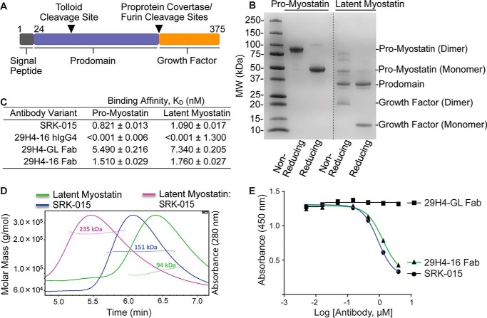
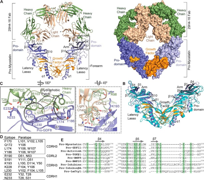
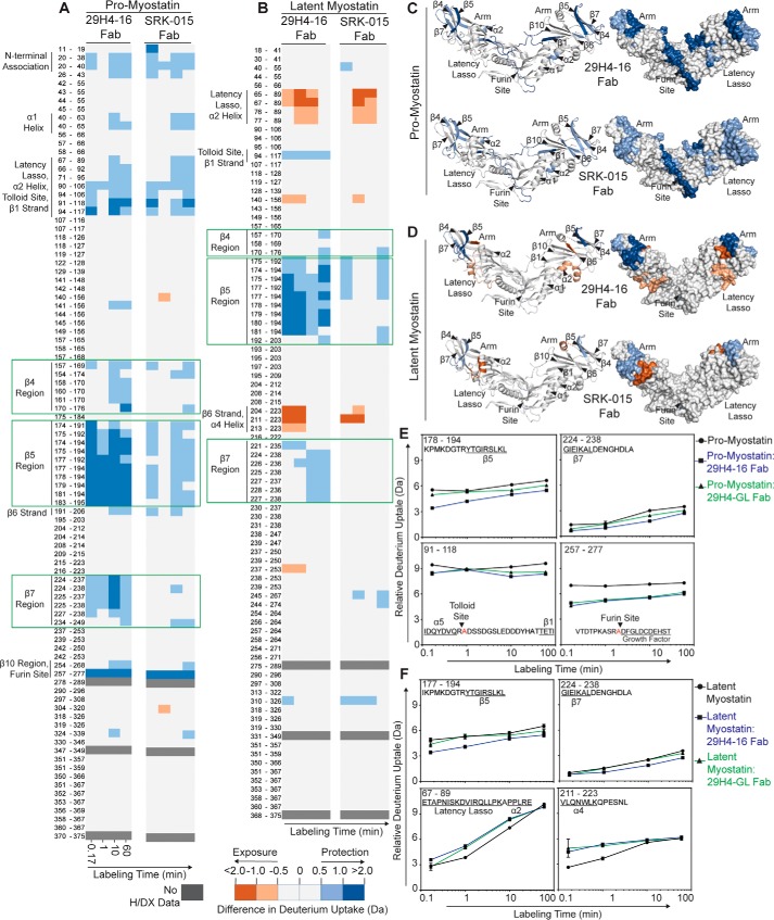
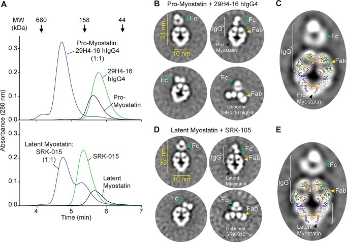
Myostatin (or growth/differentiation factor 8 (GDF8)) is a member of the transforming growth factor β superfamily of growth factors and negatively regulates skeletal muscle growth. Its dysregulation is implicated in muscle wasting diseases. SRK-015 is a clinical-stage mAb that prevents extracellular proteolytic activation of pro- and latent myostatin. Here we used integrated structural and biochemical approaches to elucidate the molecular mechanism of antibody-mediated neutralization of pro-myostatin activation. The crystal structure of pro-myostatin in complex with 29H4-16 Fab, a high-affinity variant of SRK-015, at 2.79 Å resolution revealed that the antibody binds to a conformational epitope in the arm region of the prodomain distant from the proteolytic cleavage sites. This epitope is highly sequence-divergent, having only limited similarity to other closely related members of the transforming growth factor β superfamily. Hydrogen/deuterium exchange MS experiments indicated that antibody binding induces conformational changes in pro- and latent myostatin that span the arm region, the loops contiguous to the protease cleavage sites, and the latency-associated structural elements. Moreover, negative-stain EM with full-length antibodies disclosed a stable, ring-like antigen-antibody structure in which the two Fab arms of a single antibody occupy the two arm regions of the prodomain in the pro- and latent myostatin homodimers, suggesting a 1:1 (antibody:myostatin homodimer) binding stoichiometry. These results suggest that SRK-015 binding stabilizes the latent conformation and limits the accessibility of protease cleavage sites within the prodomain. These findings shed light on approaches that specifically block the extracellular activation of growth factors by targeting their precursor forms.

Keywords: X-ray crystallography; cell signaling; growth differentiation factor 8 (GDF8); hydrogen exchange mass spectrometry; inhibition mechanism; monoclonal antibody; muscle atrophy; myostatin; proteolysis; skeletal muscle; transforming growth factor β (TGF-β).


Conflict of interest statement

All authors are current or former employees of Scholar Rock, Inc. and may be stock and/or stock option holders. G.C. and J.J. are named inventors on patents or patent applications related to SRK-015

Figures

Figure 1.
Figure 1.
SRK-015 and analogs bind and block proteolytic activation of myostatin. A, linear cartoon of pro-myostatin showing the signal peptide (black), prodomain (blue), and growth factor (orange). The tolloid and furin protease cleavage sites are indicated by inverted triangles. B, profile of Expi293F-expressed and purified pro- and latent myostatin run under nonreducing and reducing SDS-PAGE. MW, molecular weight. C, relative binding affinities of SRK-015 and analogs to pro- and latent myostatin, determined using biolayer interferometry (FortéBio Octet). Data are presented as mean ± S.E. of duplicate measurements. D, SEC-MALS profiles of latent myostatin, SRK-015, and the latent myostatin–SRK-015 complex. The dotted lines across the peaks represent the molar mass of the samples determined from the light scattering data. E, ELISA-based myostatin activity assay showing that SRK-015 and analogs block proteolytic activation of myostatin. The data are representative of three independent experiments.
Figure 2.
Figure 2.
Cocrystal structure of pro-myostatin–29H4-16 Fab. A, the overall structure of the pro- myostatin–29H4-16 Fab complex, showing two 29H4-16 Fabs occupying the arm regions in the prodomain of the pro-myostatin homodimer. The structure of the complex is shown in ribbon (left panel) and surface (right panel) representations. B, structural alignment of the 29H4-16 Fab–bound pro-myostatin structure (similar color scheme as in A) with the previously reported crystal structure of unbound pro-myostatin (cyan) (PDB code 5NTU). C, the binding interface of the pro-myostatin–29H4-16 complex, highlighting the key amino acid residues involved in the interaction. The left and right panels represent close-up views of the amino acid residues in the CDRs of the heavy (green) and light (light yellow) chains, respectively, that are in contact with the amino acid residues in the arm region of the pro-myostatin homodimer. The amino acid residues that cluster as hydrophobic patches in the binding interface are circled. D, summary of amino acid residues in the epitope and paratope at the binding interface of the pro-myostatin–29H4-16 Fab complex. E, sequence alignment of the regions (β4, β5, and β7) that covered the conformational epitope in pro-myostatin across related members of the TGF-β superfamily. Highlighted in green are the amino acid residues comprising the epitope in pro-myostatin.
Figure 3.
Figure 3.
Fluctuations in conformational dynamics of pro- and latent myostatin upon binding to antibody fragments of SRK-015 and 29H4-16 probed by H/DX-MS. A and B, heatmap showing the differences in deuterium uptake of peptic peptides identified in pro-myostatin (A) and latent myostatin (B) between its unbound and Fab-bound (SRK-015 Fab or 29H4-16 Fab) states at the indicated time of exposure to deuterated solvent. The amino acid residues for each peptic peptide are listed, and the identities of the structural elements that showed significant difference in H/D exchange between the unbound and Fab-bound states of pro- and latent myostatin are indicated. The peptic peptides covering the epitope regions are highlighted by green boxes. C and D, the significant difference in H/D exchange profiles between the unbound and Fab-bound states of pro-myostatin (C) and latent myostatin (D) after 10 min of labeling are mapped onto the model structure of pro-myostatin (PDB code 5NTU) in ribbon (left panel) and surface (right panel) representations. For these data, a deuterium uptake difference of more than 0.5 Da is considered significant at a 98% confidence interval, calculated as described previously (66). The intensities of the blue and red colors represent peptides that undergo either a significant decrease (less solvent exposure/less flexible) or increase (more solvent exposure/more flexible), respectively, during H/D exchange events between the unbound and the Fab-bound states of pro- and latent myostatin. E and F, representative deuterium incorporation plots of key peptic peptides that showed significant H/D exchange between the unbound and Fab-bound states of pro-myostatin (E) and latent myostatin (F). These peptide regions covered the epitope in the arm of the prodomain, the protease cleavage sites, and the structural elements associated with myostatin latency. Figs. S3 and S4 include all deuterium incorporation plots from these studies. Error bars, S.D. of duplicate H/DX-MS measurements done on two separate days.
Figure 4.
Figure 4.
Negative-stain EM revealed the structural features of pro- and latent myostatin in complex with 29H4-16 hIgG4 and SRK-015, respectively. A, SEC profile to confirm the complex formation of pro-myostatin–29H4-16 hIgG4 and latent myostatin–SRK-015 prior to negative-stain EM. B and D, representative EM class averages for pro-myostatin–29H4-16 hIgG4 (B) and latent myostatin–SRK-015 (D). Also observed in the class averages are unbound full-length antibodies. C and E, the coordinates from the X-ray cocrystal structure of pro-myostatin–29H4-16 Fab (Fig. 2A) showed a good fit for the density map of a representative class average of pro-myostatin:29H4-16 hIgG4 (C) and latent myostatin–SRK-15 (E). A broader set of class averages is included in Fig. S5.

References

    1. McPherron A. C., Lawler A. M., and Lee S. J. (1997) Regulation of skeletal muscle mass in mice by a new TGF-β superfamily member. Nature 387, 83–90 10.1038/387083a0 - DOI - PubMed
    1. Rodgers B. D., Interlichia J. P., Garikipati D. K., Mamidi R., Chandra M., Nelson O. L., Murry C. E., and Santana L. F. (2009) Myostatin represses physiological hypertrophy of the heart and excitation-contraction coupling. J. Physiol. 587, 4873–4886 10.1113/jphysiol.2009.172544 - DOI - PMC - PubMed
    1. Zimmers T. A., Davies M. V., Koniaris L. G., Haynes P., Esquela A. F., Tomkinson K. N., McPherron A. C., Wolfman N. M., and Lee S. J. (2002) Induction of cachexia in mice by systemically administered myostatin. Science 296, 1486–1488 10.1126/science.1069525 - DOI - PubMed
    1. Reisz-Porszasz S., Bhasin S., Artaza J. N., Shen R., Sinha-Hikim I., Hogue A., Fielder T. J., and Gonzalez-Cadavid N. F. (2003) Lower skeletal muscle mass in male transgenic mice with muscle-specific overexpression of myostatin. Am. J. Physiol. Endocrinol. Metab. 285, E876–E888 10.1152/ajpendo.00107.2003 - DOI - PubMed
    1. McPherron A. C., and Lee S. J. (1997) Double muscling in cattle due to mutations in the myostatin gene. Proc. Natl. Acad. Sci. U.S.A. 94, 12457–12461 10.1073/pnas.94.23.12457 - DOI - PMC - PubMed