. 2020 Mar 27;295(13):4252-4264.
doi: 10.1074/jbc.RA120.012767. Epub 2020 Feb 19.

The C-terminal domain of feline and bovine SAMHD1 proteins has a crucial role in lentiviral restriction

Affiliations

The C-terminal domain of feline and bovine SAMHD1 proteins has a crucial role in lentiviral restriction

Chu Wang et al. J Biol Chem. .

Abstract

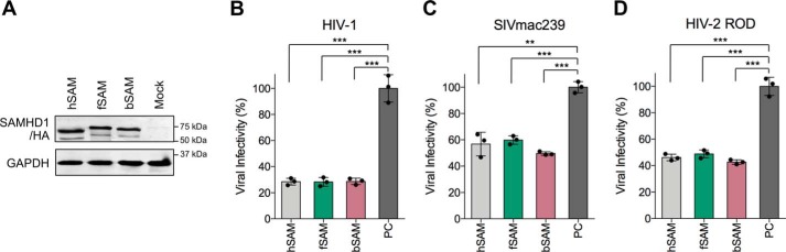
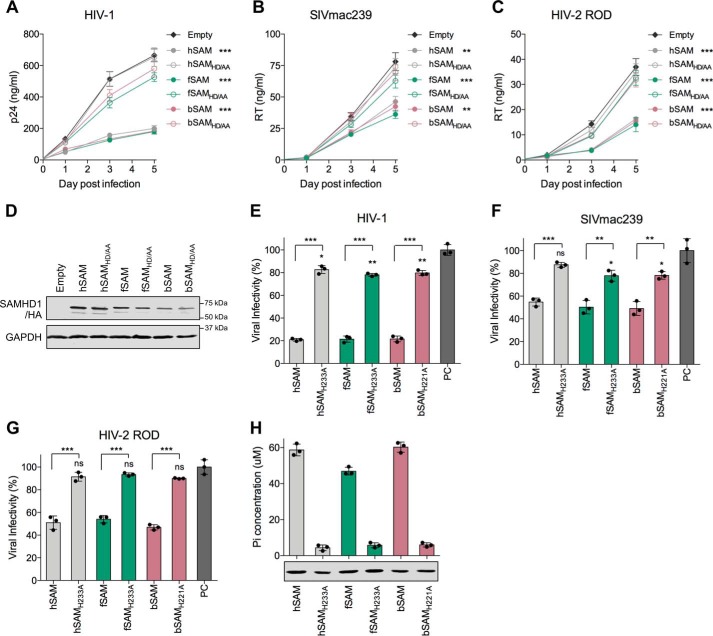
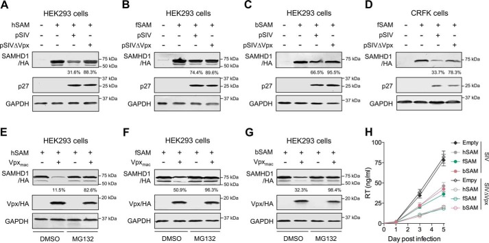
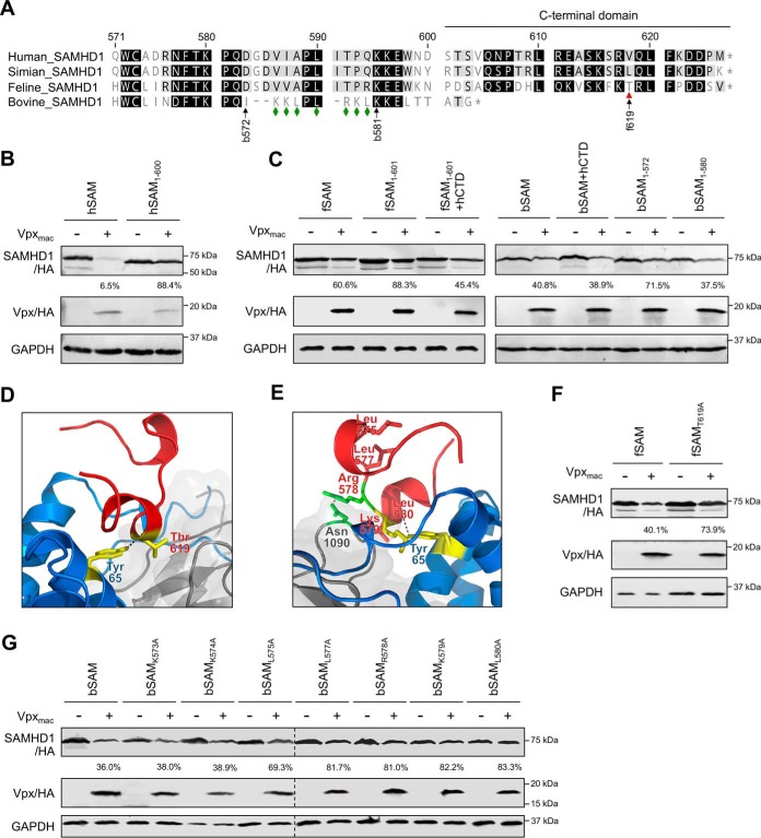
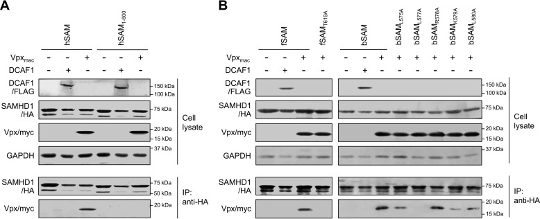
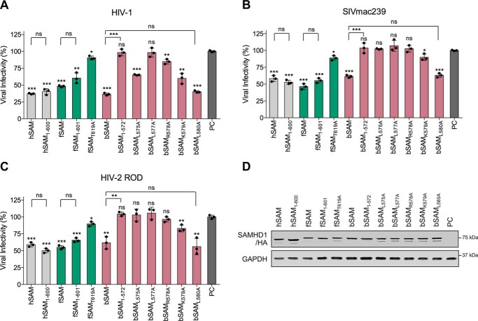
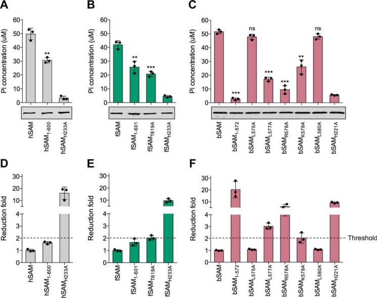
SAM and HD domain-containing protein 1 (SAMHD1) is a host factor that restricts reverse transcription of lentiviruses such as HIV in myeloid cells and resting T cells through its dNTP triphosphohydrolase (dNTPase) activity. Lentiviruses counteract this restriction by expressing the accessory protein Vpx or Vpr, which targets SAMHD1 for proteasomal degradation. SAMHD1 is conserved among mammals, and the feline and bovine SAMHD1 proteins (fSAM and bSAM) restrict lentiviruses by reducing cellular dNTP concentrations. However, the functional regions of fSAM and bSAM that are required for their biological functions are not well-characterized. Here, to establish alternative models to investigate SAMHD1 in vivo, we studied the restriction profile of fSAM and bSAM against different primate lentiviruses. We found that both fSAM and bSAM strongly restrict primate lentiviruses and that Vpx induces the proteasomal degradation of both fSAM and bSAM. Further investigation identified one and five amino acid sites in the C-terminal domain (CTD) of fSAM and bSAM, respectively, that are required for Vpx-mediated degradation. We also found that the CTD of bSAM is directly involved in mediating bSAM's antiviral activity by regulating dNTPase activity, whereas the CTD of fSAM is not. Our results suggest that the CTDs of fSAM and bSAM have important roles in their antiviral functions. These findings advance our understanding of the mechanism of fSAM- and bSAM-mediated viral restriction and might inform strategies for improving HIV animal models.

Keywords: AIDS; C-terminal domain; SAM domain and HD domain-containing protein 1 (SAMHD1); Vpx; bovine SAMHD1; dNTPase; degradation; feline SAMHD1; host-pathogen interaction; human immunodeficiency virus (HIV); lentivirus; triphosphohydrolase; viral replication; viral restriction.


Conflict of interest statement

The authors declare that they have no conflicts of interest with the contents of this article

Figures

Figure 1.
Figure 1.
The antiviral activity of feline and bovine SAMHD1 proteins against different primate lentiviruses. A, TZM-bl cells (2 × 10⁶) were transfected with 3 μg pVR1012-homo-SAMHD1-HA, pVR1012-feline-SAMHD1-HA, or pVR1012-bovine-SAMHD1-HA or empty vector (pVR1012). 48 h post transfection, the transfected cells were detached with trypsin. Partial cells of equal amounts were harvested and the expression of human, feline, and bovine SAMHD1 proteins in the transfected cells was analyzed by Western blotting using anti-HA and anti-GAPDH antibodies. B–D, the remaining cells were re-seeded into 24-well plates (2 × 10⁵ cells per well) with 300 μl viral infection solutions containing (B) HIV-1 NL4–3 (0.2 ng of RT), (C) SIVmac239 (1 ng of RT), or (D) HIV-2 ROD (1 ng of RT). 48 h later, the infected TZM-bl cells were stained with X-Gal and the positive blue cells were counted under optical microscope. Viral infectivity was determined by the number of blue cells. The viral infectivity in the empty vector–transfected cells was set to 100% (positive control, PC). Error bars represent the S.D. calculated from three independent infections. Statistical analysis was performed between the indicated groups using Student's t test.
Figure 2.
Figure 2.
dNTPase activity-dependent restriction of viral replication mediated by feline and bovine SAMHD1 proteins in U937 and TZM-bl cells. A–C, empty U937 cells (5 × 10⁵) or U937 cells stably expressing WT or catalytically inactive human, feline, or bovine SAMHD1 protein were differentiated with PMA for 20 h and infected with (A) HIV-1 NL4–3 (0.1 ng of RT), (B) SIVmac239 (1 ng of RT), or (C) HIV-2 ROD (1 ng of RT). The viral replication curves were measured by the concentration of p24 for HIV-1 or RT for SIV and HIV-2 in the culture supernatants collected on the day of infection (day 0) and at day 1, 3, and 5 post infection. Error bars represent the S.D. calculated from three independent infections. Statistical analysis was performed between each group of cells expressing WT SAMHD1 protein and the empty U937 cells using repeated-measure analysis of two-way ANOVA. D, the expression of SAMHD1 proteins in the U937 cell lines was analyzed by Western blotting using anti-HA and anti-GAPDH antibodies. E–G, TZM-bl cells (2 × 10⁶) were transfected with 3 μg pVR1012-homo-SAMHD1-HA, pVR1012-feline-SAMHD1-HA, pVR1012-bovine-SAMHD1-HA, or their mutants or empty vector and then infected with (E) HIV-1 NL4–3 (0.2 ng of RT), (F) SIVmac239 (1 ng of RT), or (G) HIV-2 ROD (1 ng of RT) at 48 h post transfection. The viral infectivity in empty vector–transfected cells was set to 100% (PC). Error bars represent the S.D. calculated from three independent infections. Statistical analysis was performed between each group of the WT proteins and their mutants and between each mutant and PC using Student's t test. H, in vitro detection of SAMHD1-catalyzed inorganic phosphate (Pi) release. HA-tagged SAMHD1 proteins were isolated from transfected HEK293 cells by immunoprecipitation. An aliquot of the immunoprecipitated SAMHD1 proteins was analyzed by Western blotting to ascertain comparable protein using anti-HA antibody. The levels of Pi released after in vitro dGTP-pyrophosphatase hydrolysis reactions were detected by malachite green. Error bars represent the S.D. calculated from three independent reactions.
Figure 3.
Figure 3.
Vpx induced degradation of feline and bovine SAMHD1 in a proteasome-dependent manner. A–C, HEK293 cells (1 × 10⁶) were co-transfected with 600 ng of (A) pVR1012-homo-SAMHD1-HA, (B) pVR1012-feline-SAMHD1-HA, or (C) pVR1012-bovine-SAMHD1-HA and 1.5 μg of pSIVmac239 or pSIVmac239ΔVpx or empty vector. D, CRFK cells (1 × 10⁶) were co-transfected with 600 ng of pVR1012-feline-SAMHD1-HA and 1.5 μg of pSIVmac239 or pSIVmac239ΔVpx or empty vector. Cells were harvested at 48 h post transfection and then analyzed by Western blotting using anti-HA, anti-SIV p27, and anti-GAPDH antibodies. The percentage of SAMHD1 in the presence of pSIVmac239 or pSIVmac239ΔVpx was calculated relative to that of the corresponding SAMHD1 in the absence of them (set to 100%). E–G, HEK293 cells (1 × 10⁶) were co-transfected with 600 ng of (E) pVR1012-homo-SAMHD1-HA, (F) pVR1012-feline-SAMHD1-HA, or (G) pVR1012-bovine-SAMHD1-HA and 500 ng of pCG-Vpxmac-HA or empty vector. The transfected cells were treated with the proteasome inhibitor MG132 at 20 μm or DMSO as a negative control at 36 h after transfection. Cells were harvested 12 h later (48 h post transfection) and then analyzed by Western blotting using anti-HA and anti-GAPDH antibodies. The percentage of SAMHD1 in the presence of Vpxmac with DMSO or MG132 treatment was calculated relative to that of the corresponding SAMHD1 in the absence of Vpxmac (set to 100%). H, empty U937 cells (5 × 10⁵) or U937 cells stably expressing each WT SAMHD1 protein were differentiated with PMA for 20 h and infected with SIVmac239 or SIVmac239ΔVpx (1 ng of RT). The viral replication curves were measured by the concentration of RT in the culture supernatants collected on the day of infection (day 0) and at day 1, 3, and 5 post infection. Error bars represent the S.D. calculated from three independent infections.
Figure 4.
Figure 4.
Vpx-induced degradation of the CTD mutants of feline and bovine SAMHD1. A, alignment of amino acids of the C-terminal region of human, simian, feline, and bovine SAMHD1 proteins. The residues in human SAMHD1 are numbered as the scale. Numbers with “f” or “b” prefix and arrows represent the position in feline or bovine SAMHD1 proteins. Mutations were made at the sites marked with red triangles and green diamonds in feline and bovine SAMHD1 sequences, respectively. B, C, F, G, HEK293 cells (1 × 10⁶) were co-transfected with 600 ng of (B) pVR1012-homo-SAMHD1-HA or (C, F, G) pVR1012-feline-SAMHD1-HA or pVR1012-bovine-SAMHD1-HA or their indicated mutants and 500 ng pCG-Vpxmac-HA or empty vector. Cell lysates were analyzed by Western blotting with anti-HA and anti-GAPDH antibodies. The dashed lines in (G) indicate the border of two spliced blots. D and E, proposed models for the interface between (D) feline SAMHD1 CTD-Vpxmac-DCAF1 CTD and (E) bovine SAMHD1 CTD-Vpxmac-DCAF1 CTD. SAMHD1 is shown in red, Vpxmac and DCAF1 CTD are in blue and gray, respectively. Residues contributing to the interface are shown as sticks, hydrogen-bonding interactions as dashed lines. Interacting residues between SAMHD1 CTD and Vpxmac are highlighted by yellow, between SAMHD1 CTD and DCAF1 CTD by green.
Figure 5.
Figure 5.
Essential amino acids of feline and bovine SAMHD1 proteins for Vpx binding. A and B, the interactions between (A) human SAMHD1 or (B) feline or bovine SAMHD1 or their indicated mutants and Vpxmac or DCAF1 were examined by co-immunoprecipitation in HEK293 cells. The transfected cells were incubated with 20 μm MG132 for 12 h before harvesting. Cell lysates were immunoprecipitated with anti-HA antibody, followed by Western blotting with anti-HA, anti-FLAG, anti-myc, and anti-GAPDH antibodies.
Figure 6.
Figure 6.
Restriction of different lentiviruses by feline and bovine SAMHD1 and their CTD mutants. A–C, TZM-bl cells (2 × 10⁶) were transfected with 3 μg pVR1012-homo-SAMHD1-HA, pVR1012-feline-SAMHD1-HA, pVR1012-bovine-SAMHD1-HA, or their indicated mutants or empty vector and then 2 × 10⁵ of the cells were infected with (A) HIV-1 (0.2 ng of RT), (B) SIVmac239 (1 ng of RT), or (C) HIV-2 ROD (1 ng of RT) at 48 h post transfection. The viral infectivity in empty vector-transfected cells was set to 100% (PC). Error bars represent the S.D. calculated from three independent infections. Statistical analysis was performed between each group and PC and between the indicated groups. D, the intracellular level of indicated SAMHD1 proteins in the transfected TZM-bl cells was analyzed by Western blotting using anti-HA and anti-GAPDH antibodies.
Figure 7.
Figure 7.
In vitro dNTPase activity of feline and bovine SAMHD1 and their CTD mutants. A–C, in vitro detection of SAMHD1-catalyzed Pi release. HA-tagged SAMHD1 proteins were isolated from transfected HEK293 cells by immunoprecipitation. An aliquot of the immunoprecipitated SAMHD1 proteins was analyzed by Western blotting to ascertain comparable protein using anti-HA antibody. The levels of Pi released after in vitro dGTP-PPase hydrolyzation reaction were detected by malachite green. Error bars represent the S.D. calculated from three independent reactions. Statistical analysis was performed between each CTD mutant and its WT SAMHD1 protein. D–F, the reduction fold was calculated by dividing the average value of Pi concentration released from dGTP-PPase hydrolyzation reaction of the WT SAMHD1 protein by each of the triplicate values from each mutant. The threshold value of Pi concentration reduction fold is 2.

Similar articles

Cited by

References

    1. Goldstone D. C., Ennis-Adeniran V., Hedden J. J., Groom H. C., Rice G. I., Christodoulou E., Walker P. A., Kelly G., Haire L. F., Yap M. W., de Carvalho L. P., Stoye J. P., Crow Y. J., Taylor I. A., and Webb M. (2011) HIV-1 restriction factor SAMHD1 is a deoxynucleoside triphosphate triphosphohydrolase. Nature 480, 379–382 10.1038/nature10623 - DOI - PubMed
    1. Powell R. D., Holland P. J., Hollis T., and Perrino F. W. (2011) Aicardi-Goutieres syndrome gene and HIV-1 restriction factor SAMHD1 is a dGTP-regulated deoxynucleotide triphosphohydrolase. J. Biol. Chem. 286, 43596–43600 10.1074/jbc.C111.317628 - DOI - PMC - PubMed
    1. Kim B., Nguyen L. A., Daddacha W., and Hollenbaugh J. A. (2012) Tight interplay among SAMHD1 protein level, cellular dNTP levels, and HIV-1 proviral DNA synthesis kinetics in human primary monocyte-derived macrophages. J. Biol. Chem. 287, 21570–21574 10.1074/jbc.C112.374843 - DOI - PMC - PubMed
    1. Lahouassa H., Daddacha W., Hofmann H., Ayinde D., Logue E. C., Dragin L., Bloch N., Maudet C., Bertrand M., Gramberg T., Pancino G., Priet S., Canard B., Laguette N., Benkirane M., Transy C., Landau N. R., Kim B., and Margottin-Goguet F. (2012) SAMHD1 restricts the replication of human immunodeficiency virus type 1 by depleting the intracellular pool of deoxynucleoside triphosphates. Nat. Immunol. 13, 223–228 10.1038/ni.2236 - DOI - PMC - PubMed
    1. St. Gelais C., de Silva S., Amie S. M., Coleman C. M., Hoy H., Hollenbaugh J. A., Kim B., and Wu L. (2012) SAMHD1 restricts HIV-1 infection in dendritic cells (DCs) by dNTP depletion, but its expression in DCs and primary CD4+ T-lymphocytes cannot be upregulated by interferons. Retrovirology 9, 105 10.1186/1742-4690-9-105 - DOI - PMC - PubMed

Publication types

Substances

Associated data